


 下载产品说明书 下载SDS
 用小程序,查商品更便捷


 收藏
收藏
 对比
对比 咨询
咨询
Scientific Data
 View Larger
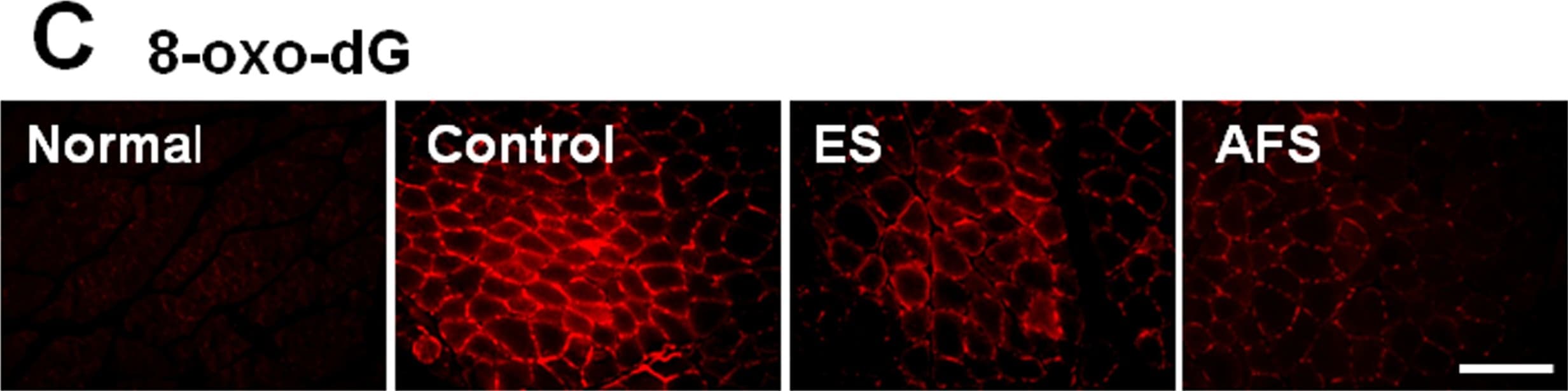
Detection of Rat 8-oxo-dG by Immunocytochemistry/ Immunofluorescence Expression of apoptotic factors in denervated muscle 2 weeks after denervation.(A) Expression of Bcl-2, Bad, and Bax in denervated muscle subjected to different treatment (B) Quantitative analysis of apoptotic markers in different treatment groups (C) The expression of 8-oxo-dG in normal, control, ES, and AFS group. (D) The representative of western blot analysis of 8-oxo-dG related to different treatment (n = 2) (F) Quantitative analysis of caspase-3 in different treatment groups Bar length = 100 μm. R = right, L = left, N = 6 for each group, * p<0.05 and **p<0.01indicated a statistical difference compared to the control group; # p<0.05 indicated a statistical difference as compared to the ES group. Image collected and cropped by CiteAb from the following open publication (https://pubmed.ncbi.nlm.nih.gov/25945496), licensed under a CC-BY license. Not internally tested by R&D Systems.
8-oxo-dG Antibody Summary
Applications
H2O2 treated MCF 10A human breast epithelial cell line
Please Note: Optimal dilutions should be determined by each laboratory for each application. General Protocols are available in the Technical Information section on our website.
Background: 8-oxo-dG
The production of 8-Hydroxyguanine (8-oxo-dG) is almost exclusively elicited by oxidative stress. Polymerases preferentially insert adenine opposite 8-oxo-dG. Therefore, oxidatively damaged adducts, without repair, are susceptible to G to T transitions.

 危险品化学品经营许可证(不带存储) 许可证编号:沪(杨)应急管危经许[2022]202944(QY)
  营业执照(三证合一)