


 用小程序,查商品更便捷


Product Usage Information
For optimal ChIP results, use 10 μl of antibody and 10 μg of chromatin (approximately 4 x 10⁶ cells) per IP. This antibody has been validated using SimpleChIP® Enzymatic Chromatin IP Kits.
| Application | Dilution |
|---|---|
| Western Blotting | 1:1000 |
| Immunoprecipitation | 1:100 |
| Immunohistochemistry (Paraffin) | 1:400 - 1:1600 |
| Immunofluorescence (Immunocytochemistry) | 1:400 - 1:1600 |
| Chromatin IP | 1:50 |
| Peptide ELISA (DELFIA) | 1:2000 |
Specificity/Sensitivity
Species Reactivity:
All Species Expected
参考图片
Western blot analysis of extracts from NIH/3T3 cells, untreated or sodium butyrate-treated (5 mM for 24 hours), showing an increase in histone acetylation using Acetylated-Lysine Antibody.
对NIH/3T3细胞抽提液,未处理或5 mM丁酸钠处理24小时,使用Acetylated-Lysine Antibody进行Western blot分析,显示组蛋白乙酰化增加。
Western blot analysis of immunoprecipitated p53 showing an increase in p53 acetylation using Acetylated-Lysine Antibody (upper) or p53 antibody (lower). p53 was immunoprecipitated from lysates from 293 cells, untreated or UV-treated, using p53 Antibody #9282.
对免疫共沉淀的p53,使用Acetylated-Lysine Antibody(上图)或p53 antibody(下图)进行Western blot分析,显示p53乙酰化增加。从293细胞裂解后免疫共沉淀的p53,未处理或UV处理,使用p53 Antibody #9282。
Immunohistochemical staining of a paraffin-embedded human breast tumor section showing nuclear and cytoplasmic localization of proteins with acetylated lysine residues using Acetylated-Lysine Antibody.
对石蜡包埋的人乳腺肿瘤切片使用Acetylated-Lysine Antibody进行免疫组化分析,显示细胞核和细胞质定位。
Western blot analysis of extracts from COS cells, untreated or TSA-treated, grown in 10% FBS (lanes 1 and 2) or serum starved for 18 hours (lanes 3 and 4), using Acetylated-Lysine Antibody (upper) or p44/42 MAP Kinase Antibody #9102 (lower).
对COS细胞,未处理或TSA处理,在10% FBS(列1和列2)生长或血清饥饿处理18小时(列3和列4),使用Acetylated-Lysine Antibody(上图)或p44/42 MAP Kinase Antibody #9102(下图)进行Western blot分析。
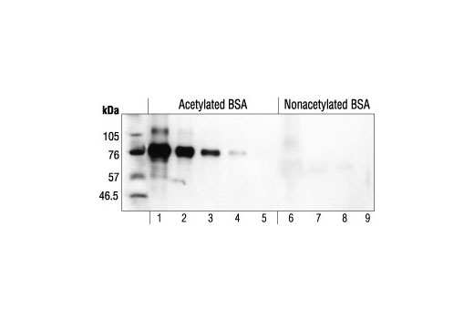
Specificity and sensitivity of Acetylated-Lysine Antibody assayed on acetylated BSA (4; 1; 0.2; 0.04 or 0.008 ng in lanes 1-5) or nonacetylated BSA (25,000; 5,000; 1,000 or 200 ng in lanes 6-9).
Acetylated-Lysine Antibody的特异性或敏感性用乙酰化BSA(4、1、0.2、0.04或0.008 ng 列1-5)和非乙酰化BSA(25,000、5,000、1,000或200 ng 列6-9)进行检测。
Immunohistochemical analysis of paraffin-embedded NIH/3T3 untreated (left) or TSA-treated (right) using Acetylated-Lysine Antibody.
对NIH/3T3细胞,未处理(左)或TSA处理(右),使用Acetylated-Lysine Antibody进行免疫组化分析。
Immunohistochemical analysis of paraffin-embedded human colon carcinoma using Acetylated-Lysine Antibody.
对石蜡包埋的人结肠癌使用Acetylated-Lysine Antibody抗体进行免疫组化分析。
Confocal immunofluorescent analysis of NIH/3T3 cells, untreated (left) or SAHA-treated (right), labeled with Acetylated-Lysine Antibody (green). Actin filaments have been labeled with Alexa Fluor R 555 phalloidin (red). Blue pseudocolor = DRAQ5® #4084 (fluorescent DNA dye).
对NIH/3T3细胞,未处理(左)或SAHA处理(右),使用Acetylated-Lysine Antibody(绿)进行共聚焦免疫荧光分析。肌动蛋白用Alexa Fluor® 555 phalloidin(红色)标记。蓝色伪彩=DRAQ5®#4084(荧光DNA染料)。
Chromatin immunoprecipitations were performed with cross-linked chromatin from 4 x 10⁶ HeLa cells and either 20 μl of Acetylated-Lysine Antibody or 2 μl of Normal Rabbit IgG #2729, using SimpleChIP® Enzymatic Chromatin IP Kit (Agarose Beads) #9002. The enriched DNA was quantified by real-time PCR, using SimpleChIP® Human GAPDH Exon 1 Primers #5516, SimpleChIP® Human RPL30 Exon 3 Primers #7014, SimpleChIP® Human MyoD1 Exon 1 Primers #4490, and SimpleChIP® Human MYT-1 Exon 1 Primers #4493. The amount of immunoprecipitated DNA in each sample is represented as signal relative to the total amount of input chromatin, which is equivalent to one.
使用SimpleChIP® Enzymatic Chromatin IP Kit (Agarose Beads) #9002试剂盒,将4 x 10^6 HeLa细胞的染色质,与20μl Acetylated-Lysine (Ac-K2-100) Rabbit mAb #9814或者2 μl of Normal Rabbit IgG #2729进行染色质免疫沉淀。使用人SimpleChIP® Human GAPDH Exon 1 Primers #5516, SimpleChIP® Human RPL30 Exon 3 Primers #7014, SimpleChIP® Human MyoD1 Exon 1 Primers #4490和SimpleChIP® Human MYT-1 Exon 1 Primers #4493进行real-time PCR用来定量富集的DNA。每个样本中免疫沉淀的DNA使用相对于输入对照中染色质的量描述,输入对照的量相当于1。

 危险品化学品经营许可证(不带存储) 许可证编号:沪(杨)应急管危经许[2022]202944(QY)
  营业执照(三证合一)