


 全部商品分类


 下载产品说明书 下载SDS
 用小程序,查商品更便捷


 收藏
收藏
 对比
对比 咨询
咨询
参考图片
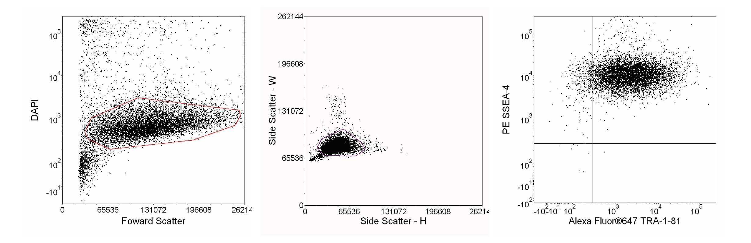
Human Embryonic Stem (ES) cells detached with Accutase™ and analyzed with cell surface markers of pluripotency. H9 ES cells (WiCell, Madison, WI) that were grown on BD Matrigel™ hESC-qualified Matrix (Cat. No. 354277) in mTeSR®1 medium (Stem Cell Technologies) were detached with Accutase™ Cell Detachment Solution. The cells were stained with either PE Mouse anti-SSEA-4 (Cat. 560128) and Alexa Fluor® 647 Mouse anti-Human TRA-1-81 (Cat. 560793) or PE Mouse IgG3, κ Isotype Control (Cat. 556659) and Alexa Fluor® 647 Mouse IgM, κ Isotype Control (Cat. 560806) and then analyzed with Diamidino-2-phenylindole dihydrochloride (DAPI) (Sigma, Cat. No. D-9542) for live/dead cell discrimination. Flow cytometry was performed on a BD LSR™ II flow cytometry system. For data analysis, live cells were first gated (left panel), and then single cells were selected by light scatter gating (center panel). Expression of surface pluripotency markers was then determined (right panel), with the positions of the quadrant markers based upon the isotype controls (data not shown).
Human Embryonic Stem (ES) cells detached with Accutase™ and analyzed with cell surface markers of pluripotency. H9 ES cells (WiCell, Madison, WI) that were grown on BD Matrigel™ hESC-qualified Matrix (Cat. No. 354277) in mTeSR®1 medium (Stem Cell Technologies) were detached with Accutase™ Cell Detachment Solution. The cells were stained with either PE Mouse anti-SSEA-4 (Cat. 560128) and Alexa Fluor® 647 Mouse anti-Human TRA-1-81 (Cat. 560793) or PE Mouse IgG3, κ Isotype Control (Cat. 556659) and Alexa Fluor® 647 Mouse IgM, κ Isotype Control (Cat. 560806) and then analyzed with Diamidino-2-phenylindole dihydrochloride (DAPI) (Sigma, Cat. No. D-9542) for live/dead cell discrimination. Flow cytometry was performed on a BD LSR™ II flow cytometry system. For data analysis, live cells were first gated (left panel), and then single cells were selected by light scatter gating (center panel). Expression of surface pluripotency markers was then determined (right panel), with the positions of the quadrant markers based upon the isotype controls (data not shown).