


 下载产品说明书
 用小程序,查商品更便捷


 收藏
收藏
 对比
对比 咨询
咨询
Specificity/Sensitivity
参考图片
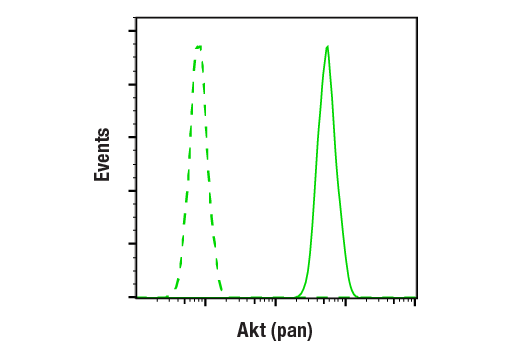
Flow cytometric analysis of Jurkat cells using Akt (pan) (C67E7) Rabbit mAb (blue) compared to a nonspecific negative control antibody (red).
Western blot analysis of recombinant Akt1, Akt2 and Akt3 proteins, and extracts from various cell lines, using Akt (pan) (C67E7) Rabbit mAb.
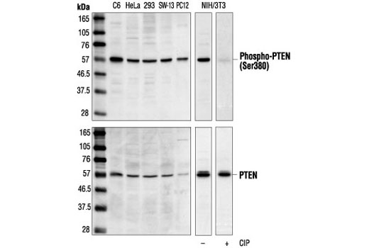
Western blot analysis of extracts from various cell lines, using Phospho-PTEN (Ser380) Antibody (upper) or PTEN Antibody #9552 (lower). The phospho-specificity of the antibody was confirmed by treating the membrane with calf intestinal alkaline phosphatase (CIP) after Western transfer.
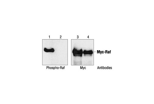
Site specificity of Phospho-c-Raf (Ser259) Antibody: Western blot analysis of recombinant Myc-tagged c-Raf protein, wild-type (lanes 1 and 3) and S259A mutant (lanes 2 and 4), using Phospho-Raf (Ser259) Antibody or a Myc antibody. (Provided by Dr. Guri Tzivion, Massachusetts General Hospital.)
After the primary antibody is bound to the target protein, a complex with HRP-linked secondary antibody is formed. The LumiGLO* is added and emits light during enzyme catalyzed decomposition.
Western blot analysis of various cell lines with Phospho-PTEN (Ser380) Antibody #9551 (upper) or PTEN Antibody #9552 (lower). The phospho-specificity of the antibody was characterized by treating the membrane without (-) or with (+) calf intestinal alkaline phosphatase (CIP) after Western transfer.
western blot分析不同细胞系,采用抗体为Phospho-PTEN (Ser380) Antibody #9551 (上) 或PTEN Antibody #9552 (下).
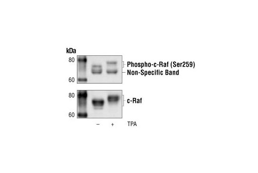
Western blot analysis of extracts from HeLa cells, untreated or TPA-treated, using Phospho-c-Raf (Ser259) Antibody (upper), or a total c-Raf antibody (lower).
Western blot analysis of extracts from HeLa cells, untreated or TPA-treated, using Phospho-Raf (Ser259) Antibody #9421 (upper), or a total c-Raf antibody (lower).
Western blot分析HeLa细胞,未处理组或TPA处理组,所用抗体为Phospho-Raf (Ser259) Antibody #9421 (上), 或a total c-Raf antibody (下).
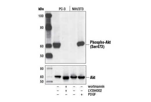
Western blot analysis of extracts from PC-3 cells, untreated or LY294002/wortmannin-treated, and NIH/3T3 cells, serum-starved or PDGF-treated, using Phospho-Akt (Ser473) (D9E) XP® Rabbit mAb (upper) or Akt (pan) (C67E7) Rabbit mAb #4691 (lower).
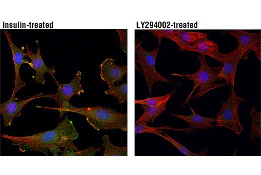
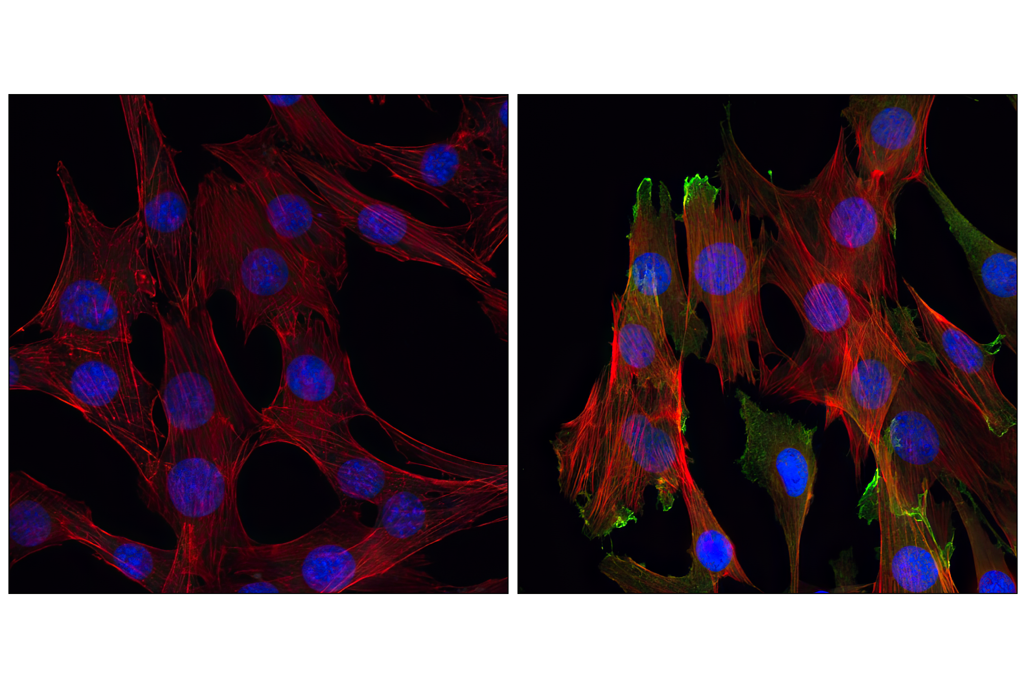
Confocal immunofluorescent analysis of C2C12 cells, LY294002-treated (left) or insulin-treated (right), using Akt (pan) (C67E7) Rabbit mAb (green). Actin filaments have been labeled with Alexa Fluor® 555 phalloidin (red). Blue pseudocolor = DRAQ5™ (fluorescent DNA dye).
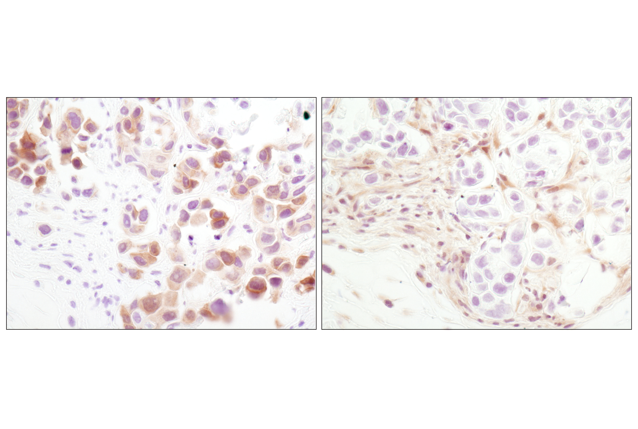
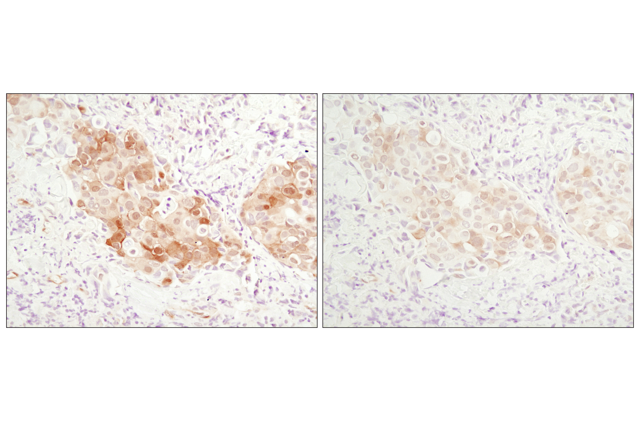
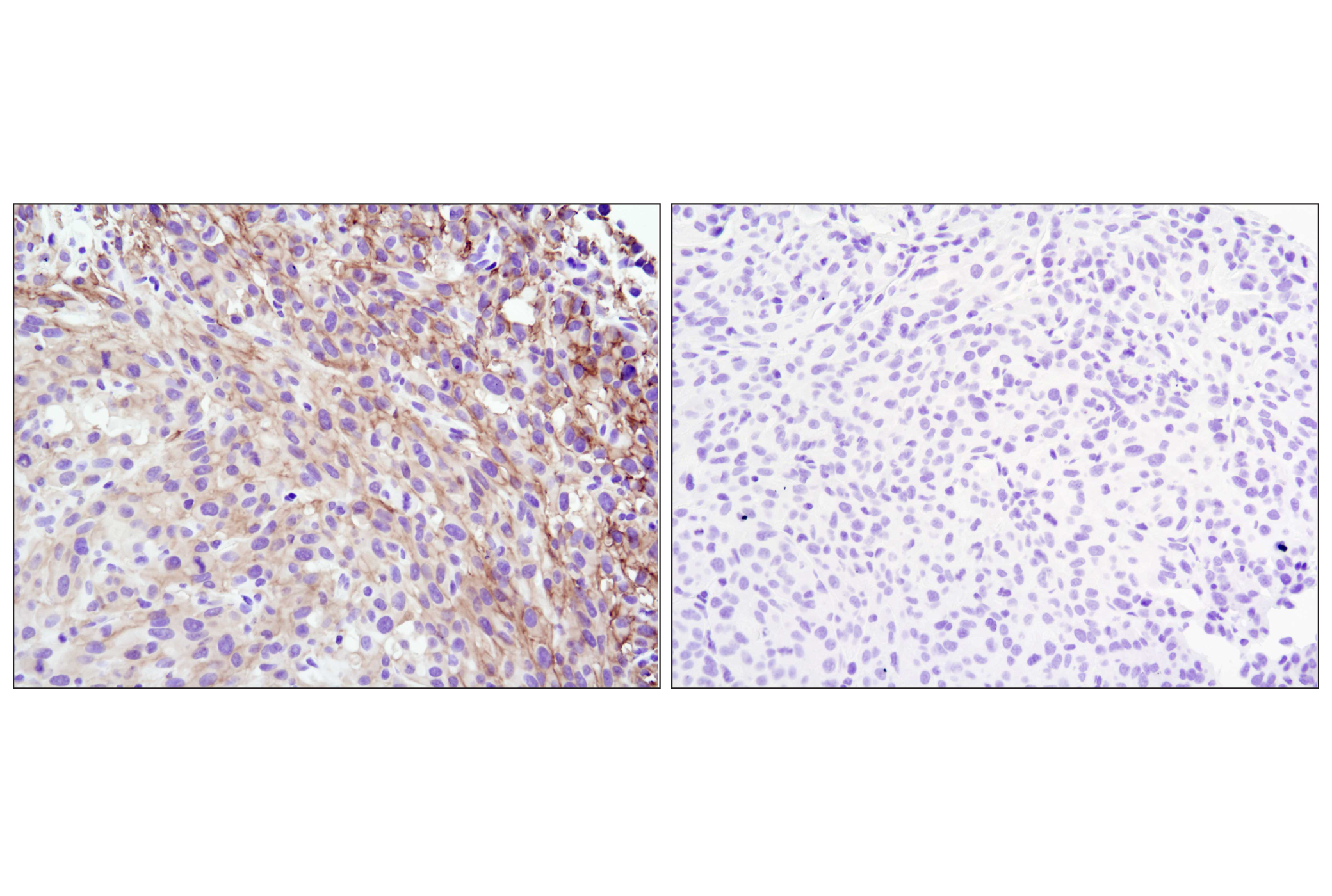
Immunohistochemical analysis of paraffin-embedded human breast carcinoma using Akt (pan) (C67E7) Rabbit mAb in the presence of control peptide (left) or Akt (pan) Blocking Peptide #1085 (right).
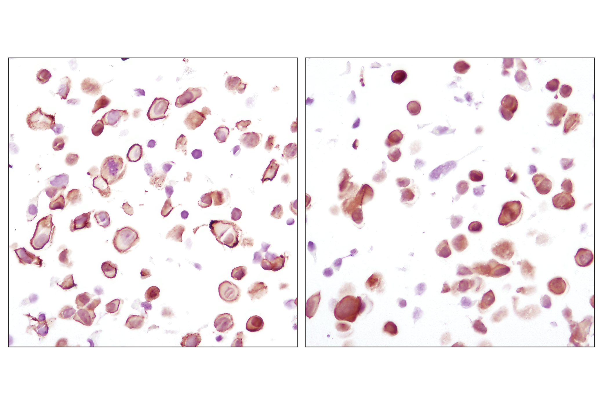
Immunohistochemical analysis using Akt (pan) (C67E7) Rabbit mAb on SignalSlide (TM) Phospho-Akt (Ser473) IHC Controls #8101 (paraffin-embedded LNCaP cells, untreated (left) or LY294002-treated (right)).
Confocal immunofluorescent analysis of C2C12 cells, LY294002-treated (left) or insulin-treated (right), using Phospho-Akt (Ser473) (D9E) XP® Rabbit mAb (green). Actin filaments have been labeled with Alexa Fluor® 555 phalloidin #8953 (red). Blue pseudocolor = DRAQ5®#4084 (fluorescent DNA dye).
Immunohistochemical analysis of paraffin-embedded U-87MG xenograft, untreated (left) or lambda phosphatase-treated (right), using Phospho-Akt (Ser473) (D9E) XP® Rabbit mAb.
Immunohistochemical analysis of paraffin-embedded MDA-MB-468 xenograft using Phospho-Akt (Ser473) (D9E) XP® Rabbit mAb (left) or PTEN (138G6) Rabbit mAb #9559 (right). Note the presence of P-Akt staining in the PTEN deficient MDA-MB-468 cells.
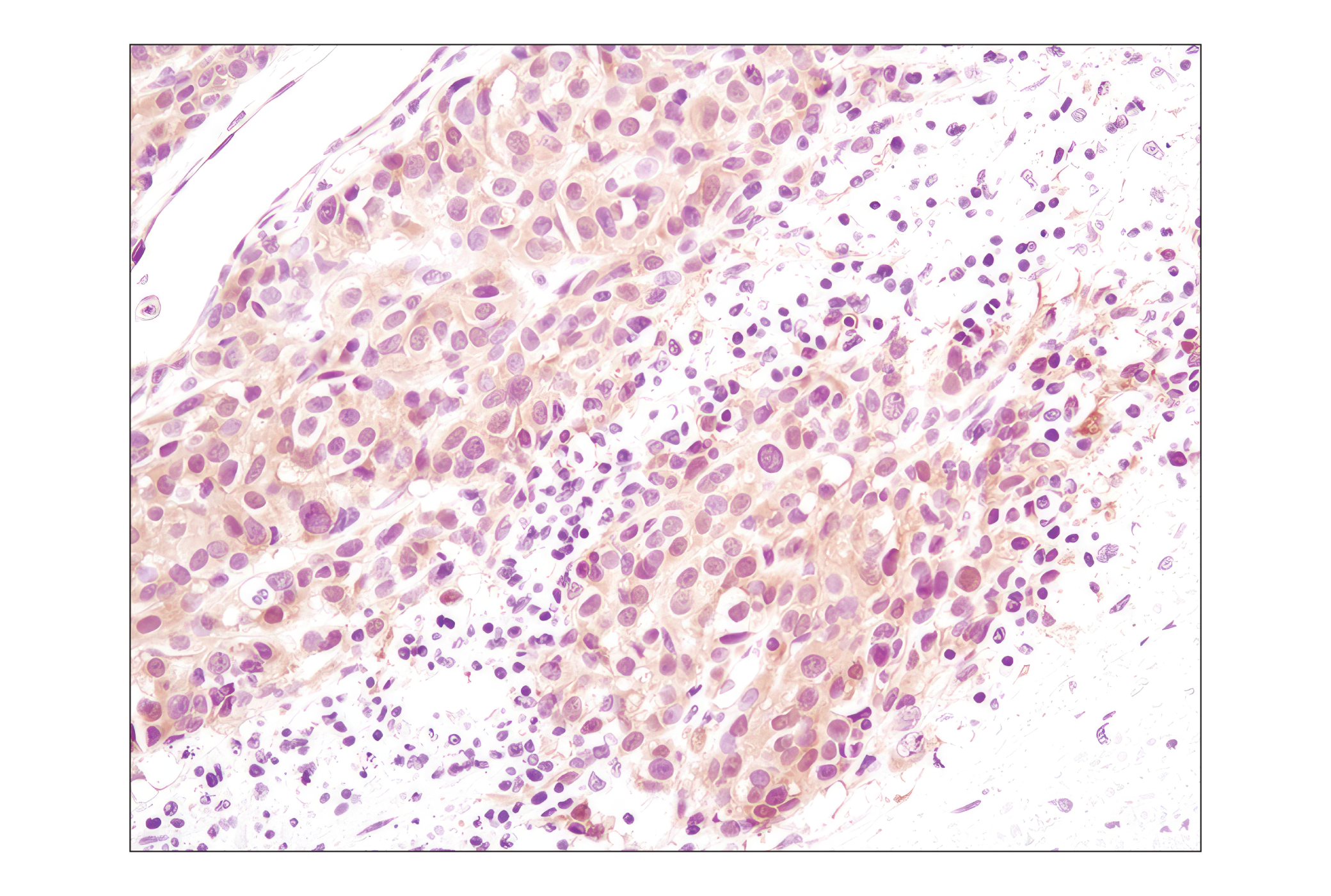
Immunohistochemical analysis of paraffin-embedded human lung carcinoma using Phospho-Akt (Ser473) (D9E) XP® Rabbit mAb.
Immunohistochemical analysis using Phospho-Akt (Ser473) (D9E) XP® Rabbit mAb on SignalSlide® Phospho-Akt (Ser473) IHC Controls #8101 (paraffin-embedded LNCaP cells, untreated (left) or LY294002-treated (right)).
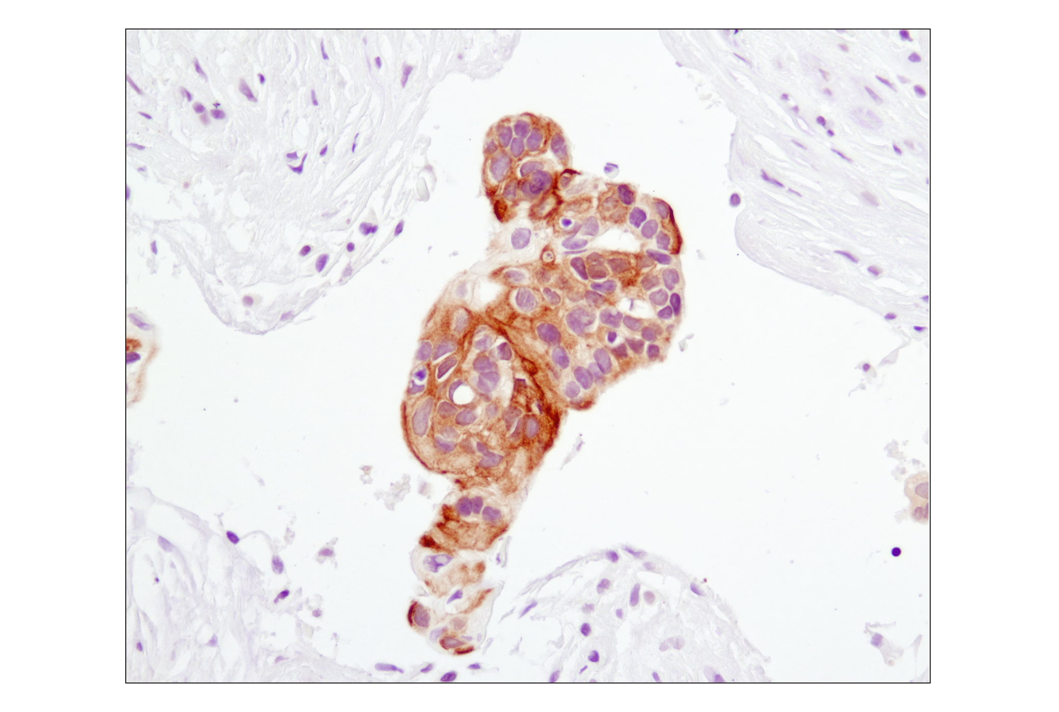
Immunohistochemical analysis of paraffin-embedded human breast carcinoma using Phospho-Akt (Ser473) (D9E) XP® Rabbit mAb.
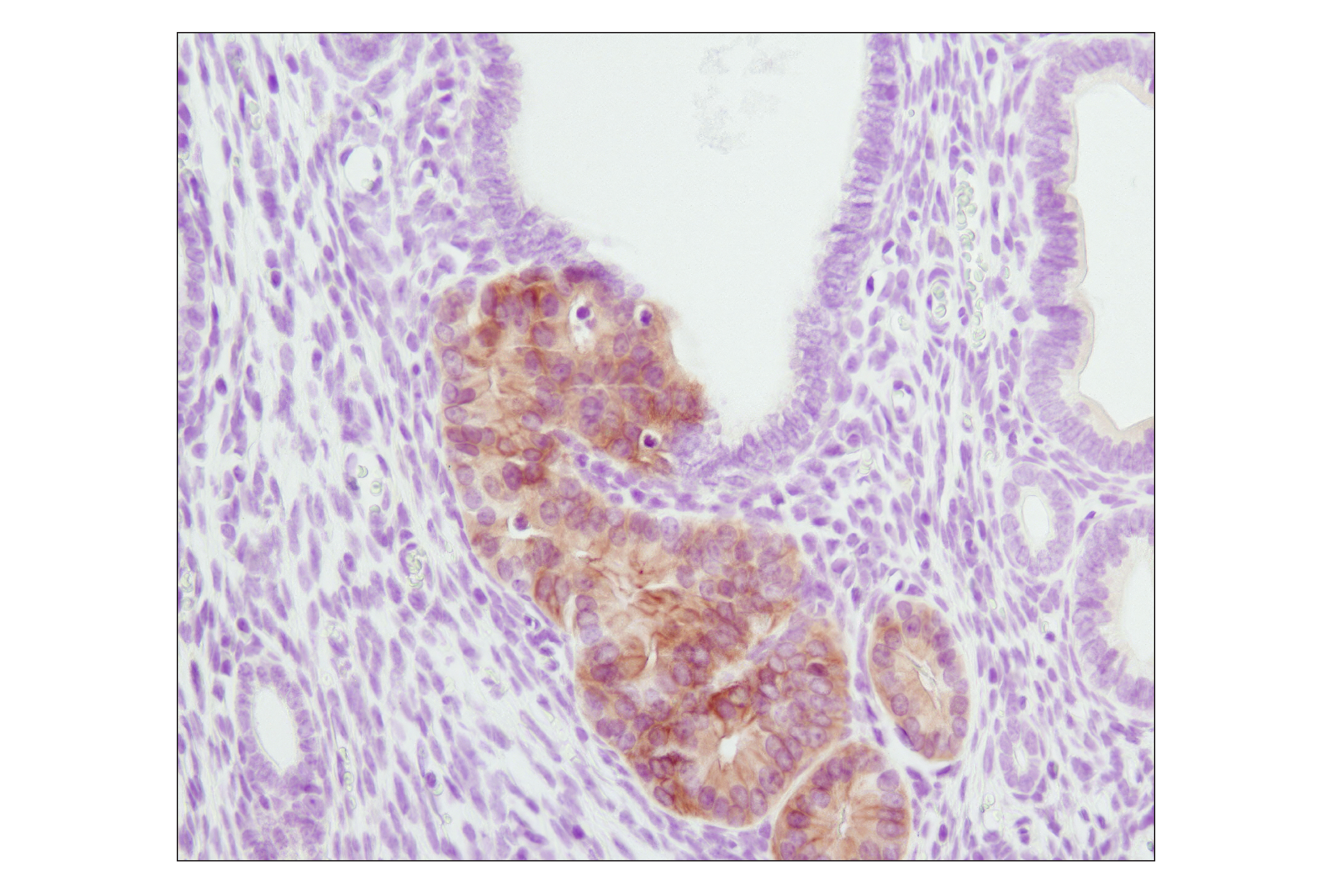
Immunohistochemical analysis of paraffin-embedded PTEN heterozygous mutant mouse endometrium using Phospho-Akt (Ser473) (D9E) XP® Rabbit mAb. (Tissue section courtesy of Dr. Sabina Signoretti, Brigham and Women's Hospital, Harvard Medical School, Boston, MA.)
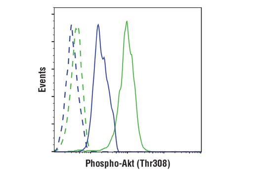
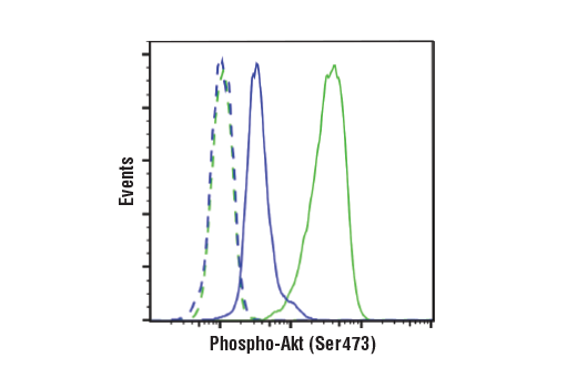
Flow cytometric analysis of Jurkat cells, untreated (green) or treated with LY294002 #9901, wortmannin #9951 and U0126 #9903 (blue), using Phospho-Akt (Ser473) (D9E) XP® Rabbit mAb compared to a nonspecific negative control antibody (red).
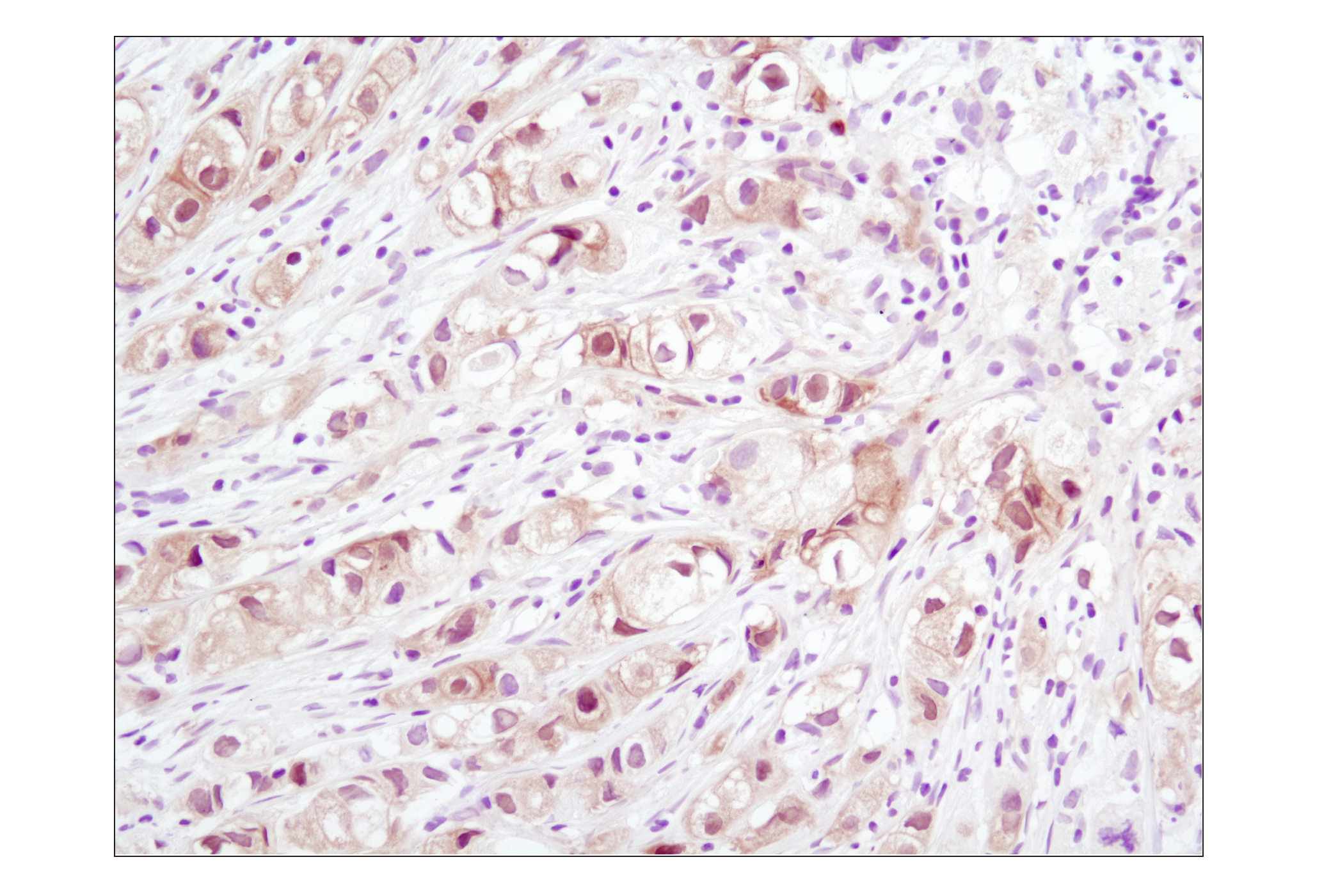
Immunohistochemical analysis of paraffin-embedded human melanoma using Akt (pan) (C67E7) Rabbit mAb.
Western blot analysis of extracts from NIH/3T3 and Jurkat cells, untreated, PDGF-treated or LY294002-treated as indicated, using Phospho-Akt (Thr308) (C31E5) Rabbit mAb #2965 (upper) or Akt (pan) (C67E7) Rabbit mAb #4691 (lower). western blot分析NIH/3T3 和 Jurkat 细胞提取物,未处理组,PDGF或LY294002处理组,所用抗体为Phospho-Akt (Thr308) (C31E5) Rabbit mAb #2965 (上) 或 Akt (pan) (C67E7) Rabbit mAb #4691 (下).
Western blot analysis of extracts from PC3 cells, untreated or LY294002/wortmannin-treated, and NIH/3T3 cells, serum- starved or PDGF-treated, using Phospho-Akt (Ser473) (D9E) XP® Rabbit mAb #4060.western blot分析PC3细胞提取物,未处理组或LY294002/wortmannin处理组和NIH/3T3,血清饥饿或PDGF处理组,所用抗体为Phospho-Akt (Ser473) (D9E) XP® Rabbit mAb #4060。
Immunohistochemical analysis of paraffin-embedded human breast carcinoma comparing SignalStain® Antibody Diluent #8112 (left) to TBST/5% normal goat serum (right) using Phospho-Akt (Ser473) (D9E) XP® Rabbit mAb #4060.
Immunohistochemical analysis of frozen SKOV3 xenograft using Phospho-Akt (Ser473) (D9E) XP® Rabbit mAb.
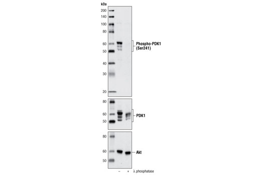
Western blot analysis of extracts from PC3 cells, untreated or λ phosphatase-treated, using Phospho-PDK1 (Ser241) (C49H2) Rabbit mAb (upper), PDK1 Antibody #3062 (middle) or Akt Antibody #9272 (lower).
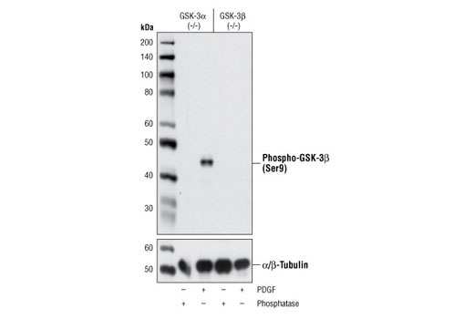
Western blot analysis of extracts from wild type (lanes 1,2), GSK-3α (-/-) (lanes 3,4) and GSK-3β (-/-) (lanes 5,6) mouse embryonic fibroblast cells (MEF), untreated or PDGF treated, using Phospho-GSK-3β (Ser9) (5B3) Rabbit mAb #9323 (upper) and GSK-3α/β Antibody (lower). (MEF wild type, GSK-3α (-/-) and GSK-3β (-/-) cells were kindly provided by Dr. Jim Woodgett, University of Toronto, Canada).
Western blot分析wild type (泳道 1,2), GSK-3α (-/-) (泳道 3,4) 和 GSK-3β (-/-) (泳道 5,6) 小鼠胚胎成纤维细胞(MEF), 未处理组或 PDGF 处理组,所用抗体为Phospho-GSK-3β (Ser9) (5B3) Rabbit mAb 兔单抗#9323 (上) 和 GSK-3α/β Antibody (下) (MEF wild type, GSK-3α (-/-) and GSK-3β (-/-) 细胞是由加拿大多伦多大学 by Dr. Jim Woodgett友情赠送。
Western blot analysis of extracts from PC3 cells, untreated or λ phosphatase-treated, using Phospho-PDK1 (Ser241) (C49H2) Rabbit mAb #3438 (upper), PDK1 Antibody #3062 (middle) or Akt Antibody #9272 (lower).
Western blot分析PC3细胞提取物,未处理组或λ 磷酸酶处理组,所用抗体为Phospho-PDK1 (Ser241) (C49H2) Rabbit mAb #3438兔单抗 (上), PDK1 Antibody #3062 (中) 或 Akt Antibody #9272 (下).
Confocal immunofluorescent analysis of wild type mouse embryonic fibroblasts (MEFs) (top row), GSK-3β (-/-) MEFs (middle row) , or PC-3 cells (bottom row), untreated (left), LY294002- and Wortmannin-treated (#9901 and #9951 respectively; center) or lambda phosphatase-treated (right), using Phospho-GSK-3β (Ser9) (D85E12) XP® Rabbit mAb (green). Actin filaments were labeled with DY-554 phalloidin (red). Blue pseudocolor = DRAQ5® #4084 (fluorescent DNA dye). (MEF wild type and GSK-3β (-/-) cells were kindly provided by Dr. Jim Woodgett, University of Toronto, Canada).
Western blot analysis of extracts from GSK-3α (-/-) (lanes 1,2) and GSK-3β (-/-) (lanes 3,4) mouse embryonic fibroblast (MEF) cells, λ phosphatase or PDGF-treated, using Phospho-GSK-3β (Ser9) (D85E12) XP® Rabbit mAb (upper) and α/β-Tubulin Antibody #2148 (lower). (MEF wild type, GSK-3α (-/-) and GSK-3β (-/-) cells were kindly provided by Dr. Jim Woodgett, University of Toronto, Canada).
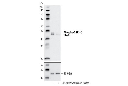
Western blot analysis of extracts from PC-3 cells, untreated or LY294002/wortmannin-treated, using Phospho-GSK-3β (Ser9) (D85E12) XP® Rabbit mAb (upper) or GSK-3β (27C10) Rabbit mAb #9315 (lower).
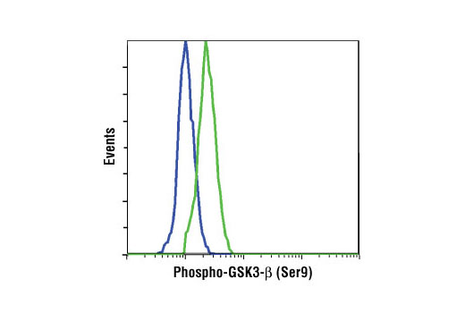
Flow cytometric analysis of NIH/3T3 cells, untreated (blue) or PDGF-treated (green), using Phospho-GSK-3β (Ser9) (D85E12) XP® Rabbit mAb.
Western blot analysis of extracts from PC3 cells, HCT116 wild-type and HCT116 PDK1 -/- cells using Phospho-PDK1 (Ser241) (C49H2) Rabbit mAb (upper) and Akt (pan) (C67E7) Rabbit mAb #4691 (lower). (HCT116 wild-type and HCT116 PDK1 -/- cells were kindly provided by Dr. Bert Vogelstein, Johns Hopkins University, Baltimore, MD).
Western blot analysis of extracts from GSK-3α (-/-) (lanes 1,2) and GSK-3β (-/-) (lanes 3,4) mouse embryonic fibroblast (MEF) cells, λ phosphatase or PDGF-treated, using Phospho-GSK-3β (Ser9) (D85E12) XP® Rabbit mAb #5558 (upper) and α/β-Tubulin Antibody #2148 (lower). (MEF wild type, GSK-3α (-/-) and GSK-3β (-/-) cells were kindly provided by Dr. Jim Woodgett, University of Toronto, Canada).
Flow cytometric analysis of serum-starved NIH/3T3 cells, untreated (blue) or treated with Human Platelet-Derived Growth Factor AA (hPDGF-AA) #8913 (100 ng/ml, 15 min; green), using Phospho-Akt (Thr308) (D25E6) XP® Rabbit mAb. Anti-rabbit IgG (H+L), F(ab')2 Fragment (Alexa Fluor® 647 Conjugate) #4414 was used as a secondary antibody.
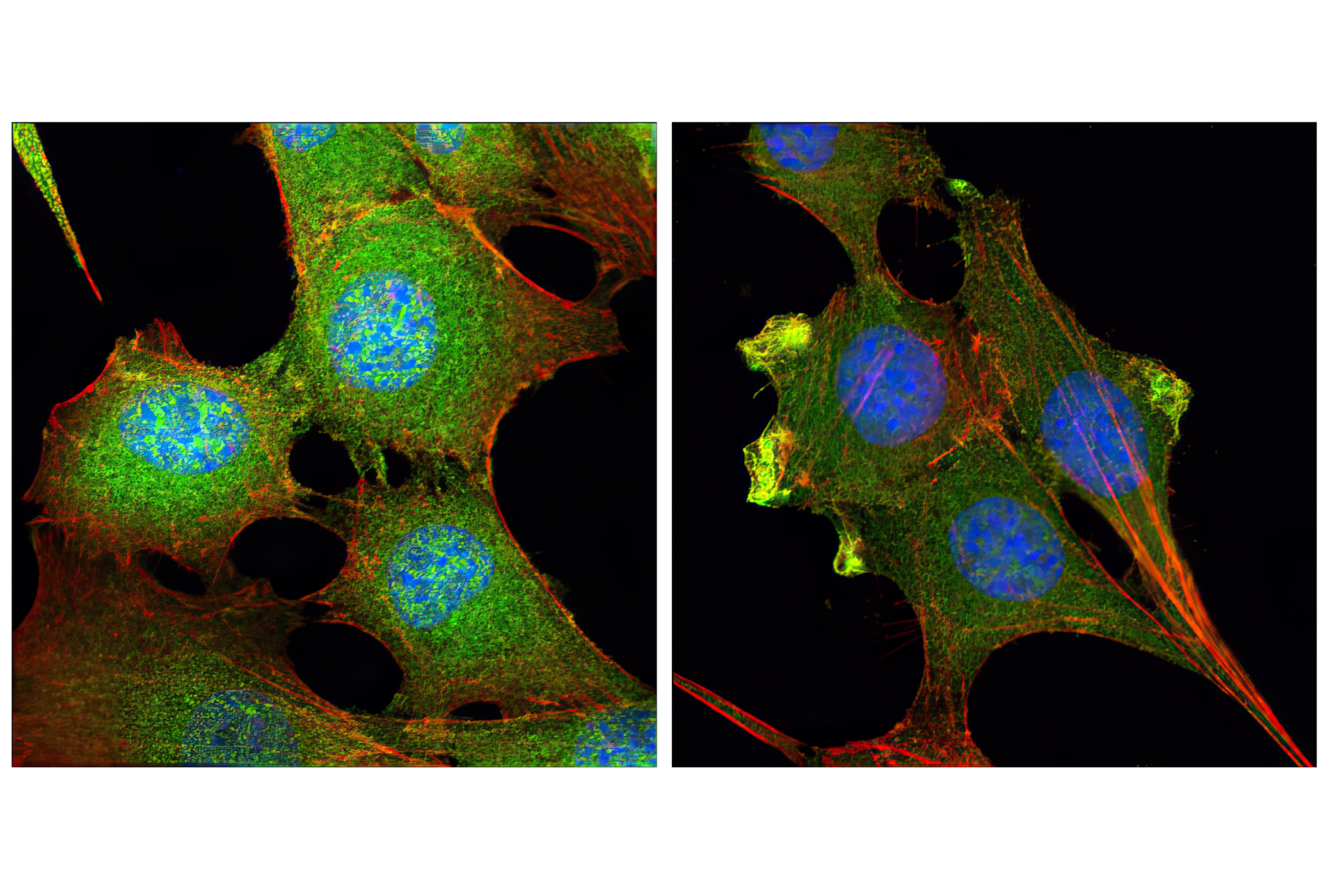
Confocal immunofluorescent analysis of C2C12 cells, insulin-treated (100 nM, 15 min; left) or treated with LY294002 #9901 (50 μM, 2 hr; right), using Phospho-Akt (Thr308) (D25E6) XP® Rabbit mAb (green). Actin filaments were labeled with DY-554 phalloidin (red). Blue pseudocolor = DRAQ5® #4084 (fluorescent DNA dye).
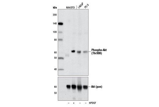
Western blot analysis of extracts from NIH/3T3 cells, untreated (-) or treated with Human Platelet-Derived Growth Factor AA (hPDGF-AA) #8913 (100 ng/ml, 5 min; +), and untreated (-) LNCaP and PC-3 cells, using Phospho-Akt (Thr308) (D25E6) XP® Rabbit mAb (upper) or Akt (pan) (C67E7) Rabbit mAb #4691 (lower).
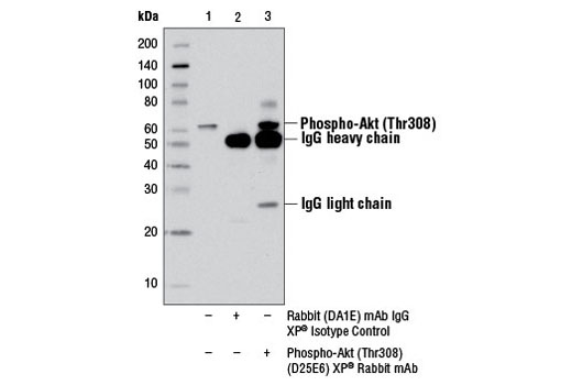
Immunoprecipitation of phospho-Akt (Thr308) from Jurkat cell extracts using Rabbit (DA1E) mAb IgG XP® Isotype Control #3900 (lane 2) or Phospho-Akt (Thr308) (D25E6) XP® Rabbit mAb (lane 3). Lane 1 is 10% input. Western blot analysis was performed using Phospho-Akt (Thr308) (D25E6) XP® Rabbit mAb.

 危险品化学品经营许可证(不带存储) 许可证编号:沪(杨)应急管危经许[2022]202944(QY)
  营业执照(三证合一)