


 下载产品说明书 下载SDS
 用小程序,查商品更便捷


 收藏
收藏
 对比
对比 咨询
咨询Scientific Data
 View Larger
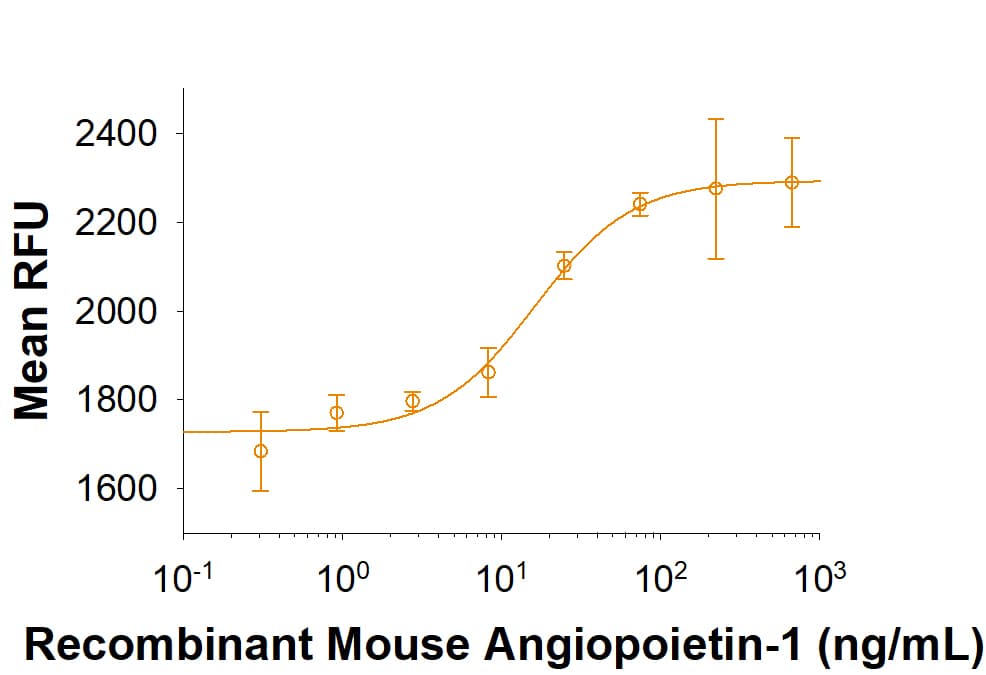
Recombinant Mouse Angiopoietin-1 inhibits serum deprivation induced apoptosis in HUVEC human umbilical vein endothelial cells. The ED50 for this effect is 10-50 ng/mL in the presence of 5 µg/mL of a cross-linking antibody, Mouse Anti-polyHistidine Monoclonal Antibody (Catalog # MAB050).
 View Larger
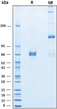
2 μg/lane of Recombinant Mouse Angiopoietin‑1 was resolved with SDS-PAGE under reducing (R) and non-reducing (NR) conditions and visualized by Coomassie® blue staining, showing bands at 59-75 kDa and oligomer, respectively.
Carrier Free
CF stands for Carrier Free (CF). We typically add Bovine Serum Albumin (BSA) as a carrier protein to our recombinant proteins. Adding a carrier protein enhances protein stability, increases shelf-life, and allows the recombinant protein to be stored at a more dilute concentration. The carrier free version does not contain BSA.
In general, we advise purchasing the recombinant protein with BSA for use in cell or tissue culture, or as an ELISA standard. In contrast, the carrier free protein is recommended for applications, in which the presence of BSA could interfere.
9936-AN
| Formulation | Lyophilized from a 0.2 μm filtered solution in Tris-Citrate and NaCl with Trehalose and with BSA as a carrier protein. |
| Reconstitution | Reconstitute at 250 μg/mL in water. |
| Shipping | The product is shipped at ambient temperature. Upon receipt, store it immediately at the temperature recommended below. |
| Stability & Storage: |
|
9936-AN/CF
| Formulation | Lyophilized from a 0.2 μm filtered solution in Tris-Citrate and NaCl with Trehalose. |
| Reconstitution | Reconstitute at 250 μg/mL in water. |
| Shipping | The product is shipped at ambient temperature. Upon receipt, store it immediately at the temperature recommended below. |
| Stability & Storage: |
|
Recombinant Mouse Angiopoietin-1 Protein Summary
Product Specifications
| Mouse Angiopoietin-1 (Ser20-Leu261) Accession # O08538 | GGGSGGGSGGGS | Mouse Angiopoietin-1 (Arg277-Phe498) Accession # O08538 | HHHHHH |
| N-terminus | C-terminus | ||
Analysis
Background: Angiopoietin-1
Angiopoietin-1 (Ang-1) is a secreted glycoprotein that plays a critical role in the development and maintenance of the vascular system (1, 2). It contains a N-terminal coiled-coil region and a C-terminal fibrinogen-like domain separated by a short flexible region (3, 4). Mature Mouse Angiopoietin-1 shares 97% and 98% amino acid sequence identity with Human and rat Angiopoietin-1, respectively. It is expressed by vascular smooth muscle cells and pericytes as an approximately 70 kDa molecule that associates into disulfide-linked homotrimers, tetramers, and pentamers (3, 5). Angiopoietin-1 binds and activates the receptor tyrosine kinase Tie-2, and its association into tetramers is important for full Tie-2 activation (3, 4). Angiopoietin-1 ligation of Tie-2 on vascular endothelial cells (EC) induces the development and branching of blood vessels (6, 7). In sub-confluent EC (i.e. during angiogenesis), Angiopoietin-1 promotes EC motility and Tie-2 localization at the trailing edge of the cell (8). In confluent EC (i.e. in homeostasis), multimeric Angiopoietin-1 enhances vascular integrity by promoting the in trans homotypic association of Tie-2 between EC or with the substratum (8, 9). In addition, Angiopoietin-1 suppresses several VEGF-induced effects on the vasculature including endothelial permeability, stretch-induced release of Angiopoietin-2, and up-regulation of the leukocyte adhesion molecules VCAM-1, ICAM-1, and E-Selectin (10-12). Angiopoietin-1 also interacts with a variety of integrins and the extracellular matrix independently of Tie-2 (13, 14). These interactions support the adhesion, migration and stress resistance of EC, fibroblasts, and myocytes (13, 14). Angiopoietin-1 can protect against pulmonary arterial hypertension (5), reduce the extent of fibrosis and remodeling in infarcted diabetic myocardium (15), and enhance tumor progression and metastasis (16).
- Koh, G.Y. (2012) Trends Mol. Med. 19:31.
- Suri, C. et al. (1996) Cell 87:1171.
- Davis, S. et al. (1996) Cell 87:1161.
- Kim, K.-T. et al. (2005) J. Biol. Chem. 280:20126.
- Kugathasan, L. et al. (2009) J. Exp. Med. 206:2221.
- Suri, C. et al. (1998) Science 282:468.
- Jeansson, M. et al. (2011) J. Clin. Invest. 121:2278.
- Saharinen, P. et al. (2008) Nat. Cell. Biol. 10:527.
- Fukuhara, S. et al. (2008) Nat. Cell Biol. 10:513.
- Jho, D. et al. (2005) Circ. Res. 96:1282.
- Korff, T. et al. (2012) Cardiovasc. Res. 94:510.
- Kim, I. et al. (2001) Circ. Res. 89:477.
- Carlson, T.R. et al. (2001) J. Biol. Chem. 276:26516.
- Dallabrida, S.M. et al. (2005) Cardiovasc. Res. 96:e8.
- Samuel, S.M. et al. (2010) Diabetes 59:51.
- Holopainen, T. et al. (2009) Cancer Res. 69:4656.


 危险品化学品经营许可证(不带存储) 许可证编号:沪(杨)应急管危经许[2022]202944(QY)
  营业执照(三证合一)