


1/2

品牌: BD Pharmingen
 下载产品说明书 下载SDS
 用小程序,查商品更便捷


 收藏
收藏
 对比
对比 咨询
咨询实验应用:
Flow cytometry (Routinely Tested)
Flow cytometry (Routinely Tested)
产品介绍
产品信息
耦联标记
APC

抗原名称
ANNEXIN V
简单描述
Apoptosis is a normal physiologic process which occurs during embryonic development as well as in maintenence of tissue homeostasis. The apoptotic program is characterized by certain morphologic features, including loss of plasma membrane asymmetry and attachment, condensation of the cytoplasm and nucleus, and internucleosomal cleavage of DNA. Loss of plasma membrane is one of the earliest features. In apoptotic cells, the membrane phospholipid phosphatidylserine (PS) is translocated from the inner to the outer leaflet of the plasma membrane, thereby exposing PS to the external cellular environment. Annexin V is a 35-36 kDa Ca2+ dependent phospholipid-binding protein that has a high affinity for PS, and binds to cells with exposed PS. Annexin V may be conjugated to fluorochromes including APC. This format retains its high affinity for PS and thus serves as a sensitive probe for flow cytometric analysis of cells that are undergoing apoptosis. Since externalization of PS occurs in the earlier stages of apoptosis, APC Annexin V staining can identify apoptosis at an earlier stage than assays based on nuclear changes such as DNA fragmentation.
APC Annexin V staining precedes the loss of membrane integrity which accompanies the latest stages of cell death resulting from either apoptotic or necrotic processes. Therefore, staining with APC Annexin V is typically used in conjunction with a vital dye such as propidium iodide (PI) or 7-Amino-Actinomycin (7-AAD) to allow the investigator to identify early apoptotic cells (7-AAD negative, APC Annexin V positive). Viable cells with intact membranes exclude 7-AAD, whereas the membranes of dead and damaged cells are permeable to 7-AAD. For example, cells that are considered viable are both APC Annexin V and 7-AAD negative while cells that are in early apoptosis are APC Annexin V positive and 7-AAD negative, while cells that are in late apoptosis or already dead are both APC Annexin V and 7-AAD positive. This assay does not distinguish between cells that have undergone apoptotic death versus those that have died as a result of a necrotic pathway because in either case, the dead cells will stain with both APC Annexin V and 7-AAD. However, when apoptosis is measured over time, cells can be often tracked from APC Annexin V and 7-AAD negative (viable, or no measurable apoptosis), to APC Annexin V positive and 7-AAD negative (early apoptosis, membrane integrity is present) and finally to APC Annexin V and 7-AAD positive (end stage apoptosis and death). The movement of cells through these three stages suggests apoptosis. In contrast, a single observation indicating that cells are both APC Annexin V and 7-AAD positive, in of itself, reveals less information about the process by which the cells underwent their demise.
APC Annexin V is routinely tested by flow cytometric analysis. Other applications were tested at BD Biosciences Pharmingen during antibody development only or reported in the literature.
商品描述
Apoptosis is a normal physiologic process which occurs during embryonic development as well as in maintenence of tissue homeostasis. The apoptotic program is characterized by certain morphologic features, including loss of plasma membrane asymmetry and attachment, condensation of the cytoplasm and nucleus, and internucleosomal cleavage of DNA. Loss of plasma membrane is one of the earliest features. In apoptotic cells, the membrane phospholipid phosphatidylserine (PS) is translocated from the inner to the outer leaflet of the plasma membrane, thereby exposing PS to the external cellular environment. Annexin V is a 35-36 kDa Ca2+ dependent phospholipid-binding protein that has a high affinity for PS, and binds to cells with exposed PS. Annexin V may be conjugated to fluorochromes including APC. This format retains its high affinity for PS and thus serves as a sensitive probe for flow cytometric analysis of cells that are undergoing apoptosis. Since externalization of PS occurs in the earlier stages of apoptosis, APC Annexin V staining can identify apoptosis at an earlier stage than assays based on nuclear changes such as DNA fragmentation.
APC Annexin V staining precedes the loss of membrane integrity which accompanies the latest stages of cell death resulting from either apoptotic or necrotic processes. Therefore, staining with APC Annexin V is typically used in conjunction with a vital dye such as propidium iodide (PI) or 7-Amino-Actinomycin (7-AAD) to allow the investigator to identify early apoptotic cells (7-AAD negative, APC Annexin V positive). Viable cells with intact membranes exclude 7-AAD, whereas the membranes of dead and damaged cells are permeable to 7-AAD. For example, cells that are considered viable are both APC Annexin V and 7-AAD negative while cells that are in early apoptosis are APC Annexin V positive and 7-AAD negative, while cells that are in late apoptosis or already dead are both APC Annexin V and 7-AAD positive. This assay does not distinguish between cells that have undergone apoptotic death versus those that have died as a result of a necrotic pathway because in either case, the dead cells will stain with both APC Annexin V and 7-AAD. However, when apoptosis is measured over time, cells can be often tracked from APC Annexin V and 7-AAD negative (viable, or no measurable apoptosis), to APC Annexin V positive and 7-AAD negative (early apoptosis, membrane integrity is present) and finally to APC Annexin V and 7-AAD positive (end stage apoptosis and death). The movement of cells through these three stages suggests apoptosis. In contrast, a single observation indicating that cells are both APC Annexin V and 7-AAD positive, in of itself, reveals less information about the process by which the cells underwent their demise.
APC Annexin V is routinely tested by flow cytometric analysis. Other applications were tested at BD Biosciences Pharmingen during antibody development only or reported in the literature.
克隆号
(RUO)
产品详情
APC
Allophycocyanin (APC), is part of the BD family of phycobiliprotein dyes. This fluorochrome is a multimeric fluorescent phycobiliprotein with excitation maximum (Ex Max) of 651 nm and an emission maximum (Em Max) at 660 nm. APC is designed to be excited by the Red (627-640 nm) laser and detected using an optical filter centered near 660 nm (e.g., a 660/20 nm bandpass filter). Please ensure that your instrument’s configurations (lasers and optical filters) are appropriate for this dye.

APC
Red 627-640 nm
651 nm
660 nm
应用
实验应用
Flow cytometry (Routinely Tested)
推荐用量
5 µl
目标/特异性
Annexin V
制备和贮存
存储溶液
Aqueous buffered solution containing BSA and ≤0.09% sodium azide.
保存方式
Aqueous buffered solution containing BSA and ≤0.09% sodium azide.
文献
文献
研发参考(8)
1. Andree HA, Reutelingsperger CP, Hauptmann R, Hemker HC, Hermens WT, Willems GM. Binding of vascular anticoagulant alpha (VAC alpha) to planar phospholipid bilayers. J Biol Chem. 1990; 265(9):4923-4928. (Biology).
2. Casciola-Rosen L, Rosen A, Petri M, Schlissel M. Surface blebs on apoptotic cells are sites of enhanced procoagulant activity: implications for coagulation events and antigenic spread in systemic lupus erythematosus. Proc Natl Acad Sci U S A. 1996; 93(4):1624-1629. (Methodology: Apoptosis, Flow cytometry).
3. Homburg CH, de Haas M, von dem Borne AE, Verhoeven AJ, Reutelingsperger CP, Roos D. Human neutrophils lose their surface Fc gamma RIII and acquire Annexin V binding sites during apoptosis in vitro. Blood. 1995; 85(2):532-540. (Biology).
4. Koopman G, Reutelingsperger CP, Kuijten GA, Keehnen RM, Pals ST, van Oers MH. Annexin V for flow cytometric detection of phosphatidylserine expression on B cells undergoing apoptosis. Blood. 1994; 84(5):1415-1420. (Methodology: Apoptosis, Flow cytometry).
5. Martin SJ, Reutelingsperger CP, McGahon AJ, et al. Early redistribution of plasma membrane phosphatidylserine is a general feature of apoptosis regardless of the initiating stimulus: inhibition by overexpression of Bcl-2 and Abl. J Exp Med. 1995; 182(5):1545-1556. (Biology).
6. Raynal P, Pollard HB. Annexins: the problem of assessing the biological role for a gene family of multifunctional calcium- and phospholipid-binding proteins. Biochim Biophys Acta. 1994; 1197(1):63-93. (Biology).
7. Vermes I, Haanen C, Steffens-Nakken H, Reutelingsperger C. A novel assay for apoptosis. Flow cytometric detection of phosphatidylserine expression on early apoptotic cells using fluorescein labelled Annexin V. J Immunol Methods. 1995; 184(1):39-51. (Methodology: Apoptosis, Flow cytometry).
8. van Engeland M, Ramaekers FC, Schutte B, Reutelingsperger CP. A novel assay to measure loss of plasma membrane asymmetry during apoptosis of adherent cells in culture. Cytometry. 1996; 24(2):131-139. (Methodology: Apoptosis, Flow cytometry).
参考图片
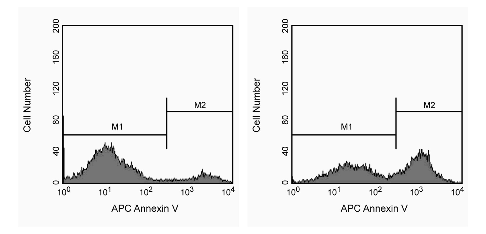
APC Annexin V: A tool for identifying cells that are undergoing apoptosis. Jurkat T cells were left untreated (left panel) or treated for 4 hours (right panel) with 6 µM camptothecin. Cells were incubated with APC Annexin V and analyzed by flow cytometry. Untreated cells were primarily APC Annexin V negative, indicating that they were viable and not undergoing apoptosis. After a 4 hour treatment with camptothecin, there were two populations of cells: cells undergoing apoptosis (APC Annexin V positive), and cells that were viable and not undergoing apoptosis (APC Annexin V negative).
声明 :本官网所有报价均为常温或者蓝冰运输价格,如有产品需要干冰运输,需另外加收干冰运输费。

 危险品化学品经营许可证(不带存储) 许可证编号:沪(杨)应急管危经许[2022]202944(QY)
  营业执照(三证合一)