


 下载产品说明书
 用小程序,查商品更便捷


 收藏
收藏
 对比
对比 咨询
咨询
- 描述数量/尺寸零件号EntrezGene ID
- Purified Recombinant Annexin V100 µg (1 ea)51-65871AN/A
- FITC Annexin V0.5 mL (1 ea)51-65874XN/A
- Propidium Iodide Staining Solution2 mL (1 ea)51-66211EN/A
- 10X Annexin V Binding Buffer50 mL (1 ea)51-66121EN/A
参考图片
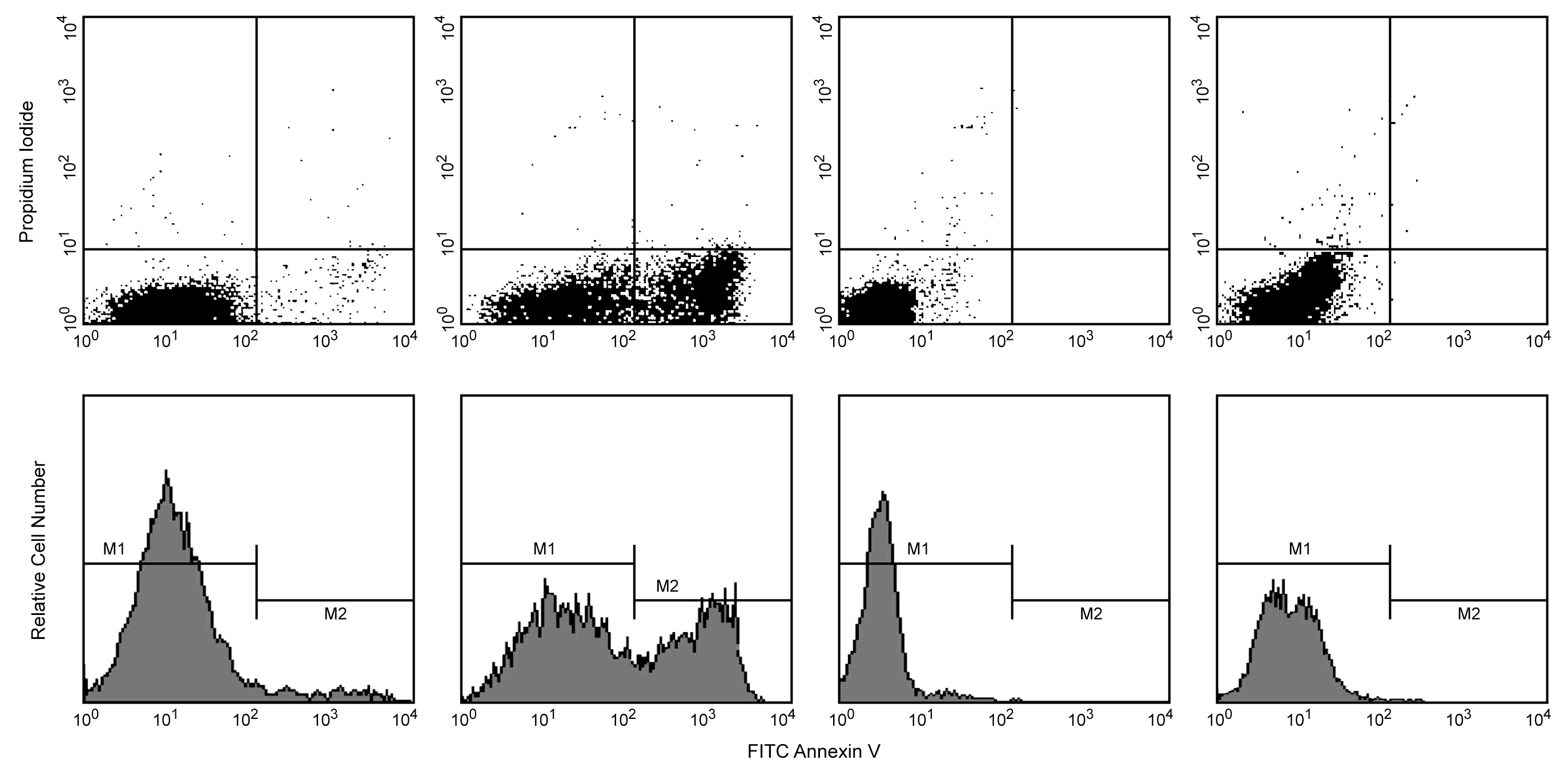
Flow Cytometric Analysis of FITC Annexin V staining and blocking. Jurkat cells (Human T-cell leukemia; ATCC TIB-152) were either untreated (top/bottom far left and middle-right panels) or were induced to undergo apoptosis by treatment with camptothecin (12 µM, 4 hours) (top/bottom middle-left and far right panels). Cells were then incubated with FITC Annexin V in a buffer containing propidium iodide (top/bottom far left and middle left panels) or with purified recombinant Annexin V (top/bottom far right and middle-right panels) in order to block FITC Annexin V binding sites prior to adding FITC Annexin-V. After camptothecin treatment (4 hours), a population of cells was FITC Annexin V positive (bottom middle-left panel, M2). FITC Annexin V staining was blocked when cells were first incubated with purified recombinant Annexin V (bottom far right panel, M2). As expected, the cell population that was not treated was primarily Annexin V negative (bottom far left panel, M1). The small number of Annexin V positive cells in the untreated population likely represents a basal level of apoptosis (bottom far left panel, M2).
Flow Cytometric Analysis of FITC Annexin V staining and blocking. Jurkat cells (Human T-cell leukemia; ATCC TIB-152) were either untreated (top/bottom far left and middle-right panels) or were induced to undergo apoptosis by treatment with camptothecin (12 µM, 4 hours) (top/bottom middle-left and far right panels). Cells were then incubated with FITC Annexin V in a buffer containing propidium iodide (top/bottom far left and middle left panels) or with purified recombinant Annexin V (top/bottom far right and middle-right panels) in order to block FITC Annexin V binding sites prior to adding FITC Annexin-V. After camptothecin treatment (4 hours), a population of cells was FITC Annexin V positive (bottom middle-left panel, M2). FITC Annexin V staining was blocked when cells were first incubated with purified recombinant Annexin V (bottom far right panel, M2). As expected, the cell population that was not treated was primarily Annexin V negative (bottom far left panel, M1). The small number of Annexin V positive cells in the untreated population likely represents a basal level of apoptosis (bottom far left panel, M2).

 危险品化学品经营许可证(不带存储) 许可证编号:沪(杨)应急管危经许[2022]202944(QY)
  营业执照(三证合一)