BD Horizon™ V450 Annexin V
1/2

BD Horizon™ V450 Annexin V

分享
品牌: BD Pharmingen
pdf 下载产品说明书
pdf 下载SDS
用小程序,查商品更便捷
收藏
对比对比
咨询咨询
    实验应用:
    Flow cytometry (Routinely Tested)
    Flow cytometry (Routinely Tested)
    产品介绍
    产品信息
    耦联标记
    V450
    抗原名称
    ANNEXIN V
    简单描述
    Apoptosis is a normal physiological process that occurs during embryonic development and tissue homeostasis maintenance. The apoptotic program is characterized by certain morphological features, including plasma membrane symmetry and detachment, condensation of the cytoplasm and nucleus, and internucleosomal cleavage of DNA. Loss of the plasma membrane is one of the earliest features of apoptosis. In apoptotic cells, phosphatidylserine (PS), a phospholipid membrane component, is translocated from the inner to the outer leaflet of the plasma membrane, thereby exposing PS to the external cellular environment. Annexin V is a 35-36 kDa Ca2+ dependent phospholipid-binding protein that has a high affinity for PS, and binds to cells with exposed PS. Annexin V may be conjugated to fluorochromes including BD Horizon™ V450. This format retains its high affinity for PS and thus serves as a sensitive probe for flow cytometric analysis of cells that are undergoing apoptosis. Since externalization of PS occurs in the earlier stages of apoptosis, V450 Annexin V staining can identify apoptosis at an earlier stage than assays based on nuclear changes such as DNA fragmentation.   V450 Annexin V staining precedes the loss of membrane integrity which accompanies the latest stages of cell death resulting from either apoptotic or necrotic processes. Therefore, staining with V450 Annexin V is typically used in conjunction with a vital dye such as propidium iodide (PI) or 7-Amino-Actinomycin (7-AAD) to allow the investigator to identify early apoptotic cells (PI negative, V450 Annexin V positive). Viable cells with intact membranes exclude PI, whereas the membranes of dead and damaged cells are permeable to PI. For example, cells that are considered viable are both V450 Annexin V and PI negative, cells that are in early apoptosis are V450 Annexin V positive and PI negative, and cells that are in late apoptosis or already dead are both V450 Annexin V and PI positive. This assay does not distinguish between cells that have undergone the apoptotic versus necrotic pathway because dead cells will be stained with both V450 Annexin V and PI. However, when apoptosis is measured over time, cells can often be tracked from V450 Annexin V and PI negative (viable, or no measurable apoptosis), to V450 Annexin V positive and PI negative (early apoptosis, membrane integrity is present) and finally to V450 Annexin V and PI positive (end-stage apoptosis and death). The movement of cells through these three stages suggests apoptosis. In contrast, a single observation indicating that cells are both V450 Annexin V and PI positive, in and of itself, reveals less information about the process by which the cells underwent their demise.   This reagent is conjugated to BD Horizon™ V450, which has been developed for use in multicolor flow cytometry experiments and is available exclusively from BD Biosciences. It is excited by the Violet laser Ex max of 406 nm and has an Em Max at 450 nm. Conjugates with BD Horizon™ V450 can be used in place of Pacific Blue™ conjugates.
    商品描述
    N/A Apoptosis is a normal physiological process that occurs during embryonic development and tissue homeostasis maintenance. The apoptotic program is characterized by certain morphological features, including plasma membrane symmetry and detachment, condensation of the cytoplasm and nucleus, and internucleosomal cleavage of DNA. Loss of the plasma membrane is one of the earliest features of apoptosis. In apoptotic cells, phosphatidylserine (PS), a phospholipid membrane component, is translocated from the inner to the outer leaflet of the plasma membrane, thereby exposing PS to the external cellular environment. Annexin V is a 35-36 kDa Ca2+ dependent phospholipid-binding protein that has a high affinity for PS, and binds to cells with exposed PS. Annexin V may be conjugated to fluorochromes including BD Horizon™ V450. This format retains its high affinity for PS and thus serves as a sensitive probe for flow cytometric analysis of cells that are undergoing apoptosis. Since externalization of PS occurs in the earlier stages of apoptosis, V450 Annexin V staining can identify apoptosis at an earlier stage than assays based on nuclear changes such as DNA fragmentation. V450 Annexin V staining precedes the loss of membrane integrity which accompanies the latest stages of cell death resulting from either apoptotic or necrotic processes. Therefore, staining with V450 Annexin V is typically used in conjunction with a vital dye such as propidium iodide (PI) or 7-Amino-Actinomycin (7-AAD) to allow the investigator to identify early apoptotic cells (PI negative, V450 Annexin V positive). Viable cells with intact membranes exclude PI, whereas the membranes of dead and damaged cells are permeable to PI. For example, cells that are considered viable are both V450 Annexin V and PI negative, cells that are in early apoptosis are V450 Annexin V positive and PI negative, and cells that are in late apoptosis or already dead are both V450 Annexin V and PI positive. This assay does not distinguish between cells that have undergone the apoptotic versus necrotic pathway because dead cells will be stained with both V450 Annexin V and PI. However, when apoptosis is measured over time, cells can often be tracked from V450 Annexin V and PI negative (viable, or no measurable apoptosis), to V450 Annexin V positive and PI negative (early apoptosis, membrane integrity is present) and finally to V450 Annexin V and PI positive (end-stage apoptosis and death). The movement of cells through these three stages suggests apoptosis. In contrast, a single observation indicating that cells are both V450 Annexin V and PI positive, in and of itself, reveals less information about the process by which the cells underwent their demise. This reagent is conjugated to BD Horizon™ V450, which has been developed for use in multicolor flow cytometry experiments and is available exclusively from BD Biosciences. It is excited by the Violet laser Ex max of 406 nm and has an Em Max at 450 nm. Conjugates with BD Horizon™ V450 can be used in place of Pacific Blue™ conjugates.
    克隆号
    克隆 N/A (RUO)
    产品详情
    V450
    BD Horizon™ V450 Dye is part of the BD Horizon™ violet family of dyes. This is a small organic fluorochrome with an excitation maximum (Ex Max) at 405-nm and an emission maximum (Em Max) at 450-nm. BD Horizon™ V450, driven by BD innovation, is designed to be excited by the violet laser (405 nm) and detected using an optical filter centered near 450-nm (e.g., a 450/50-nm bandpass filter). The dye can be excited by the UV (355-nm) laser resulting in cross-laser excitation and spillover. Please ensure that your instrument’s configurations (lasers and optical filters) are appropriate for this dye.
    V450
    Violet 405 nm
    405 nm
    450 nm
    应用
    实验应用
    Flow cytometry (Routinely Tested)
    推荐用量
    5 µl
    目标/特异性
    Annexin V
    制备和贮存
    存储溶液
    Aqueous buffered solution containing BSA, protein stabilizer, and ≤0.09% sodium azide.
    保存方式
    Aqueous buffered solution containing BSA, protein stabilizer, and ≤0.09% sodium azide.
    文献
    文献
    研发参考(12) 1. Andree HA, Reutelingsperger CP, Hauptmann R, Hemker HC, Hermens WT, Willems GM. Binding of vascular anticoagulant alpha (VAC alpha) to planar phospholipid bilayers. J Biol Chem. 1990; 265(9):4923-4928. (Biology). 2. Bossy-Wetzel E and Green D. Detection of apoptosis by Annexin V labeling. 2000; 322:15-18. (Biology). 3. Casciola-Rosen L, Rosen A, Petri M, Schlissel M. Surface blebs on apoptotic cells are sites of enhanced procoagulant activity: implications for coagulation events and antigenic spread in systemic lupus erythematosus. Proc Natl Acad Sci U S A. 1996; 93(4):1624-1629. (Methodology: Apoptosis, Flow cytometry). 4. Homburg CH, de Haas M, von dem Borne AE, Verhoeven AJ, Reutelingsperger CP, Roos D. Human neutrophils lose their surface Fc gamma RIII and acquire Annexin V binding sites during apoptosis in vitro. Blood. 1995; 85(2):532-540. (Biology). 
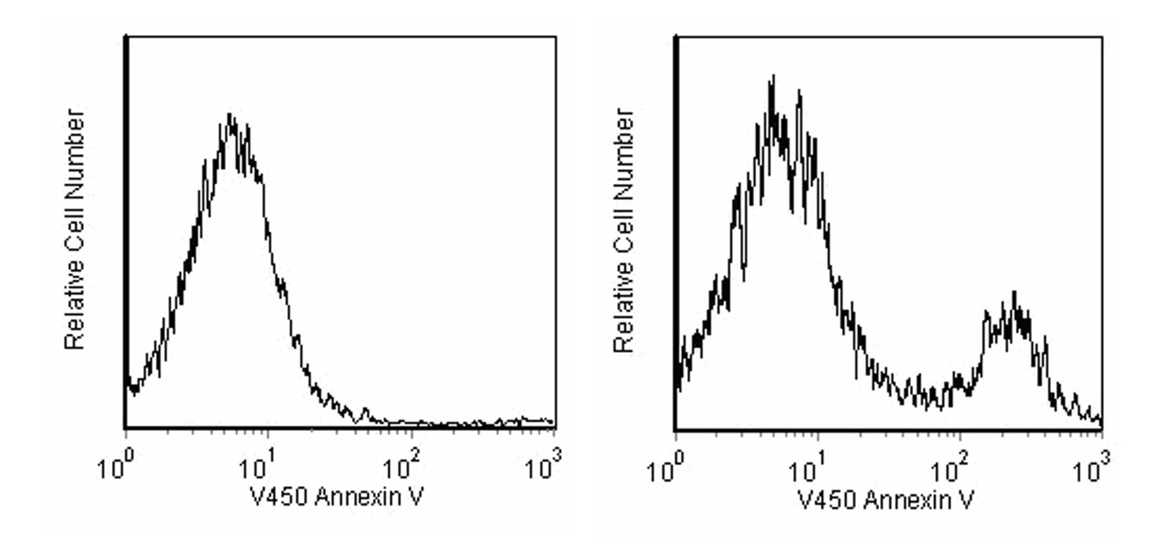
5. Koopman G, Reutelingsperger CP, Kuijten GA, Keehnen RM, Pals ST, van Oers MH. Annexin V for flow cytometric detection of phosphatidylserine expression on B cells undergoing apoptosis. Blood. 1994; 84(5):1415-1420. (Methodology: Apoptosis, Flow cytometry). 6. Martin SJ, Reutelingsperger CP, McGahon AJ, et al. Early redistribution of plasma membrane phosphatidylserine is a general feature of apoptosis regardless of the initiating stimulus: inhibition by overexpression of Bcl-2 and Abl. J Exp Med. 1995; 182(5):1545-1556. (Biology). 7. O'Brien MC, Bolton WE. Comparison of cell viability probes compatible with fixation and permeabilization for combined surface and intracellular staining in flow cytometry. Cytometry. 1995; 19(3):243-255. (Biology). 8. Raynal P, Pollard HB. Annexins: the problem of assessing the biological role for a gene family of multifunctional calcium- and phospholipid-binding proteins. Biochim Biophys Acta. 1994; 1197(1):63-93. (Biology). 9. Schmid I, Krall WJ, Uittenbogaart CH, Braun J, Giorgi JV. Dead cell discrimination with 7-amino-actinomycin D in combination with dual color immunofluorescence in single laser flow cytometry. Cytometry. 1992; 13(2):204-208. (Biology). 10. Van Engeland, Nieland LJW, Ramaekers FCS, Schutte B, and Reutelingsperger CPM. . Annexin V-affinity assay: A review on an apoptosis detection system based on phosphatidylserine exposure. Cytometry. 1998; 31:1-9. (Biology). 11. Vermes I, Haanen C, Steffens-Nakken H, Reutelingsperger C. A novel assay for apoptosis. Flow cytometric detection of phosphatidylserine expression on early apoptotic cells using fluorescein labelled Annexin V. J Immunol Methods. 1995; 184(1):39-51. (Methodology: Apoptosis, Flow cytometry). 12. van Engeland M, Ramaekers FC, Schutte B, Reutelingsperger CP. A novel assay to measure loss of plasma membrane asymmetry during apoptosis of adherent cells in culture. Cytometry. 1996; 24(2):131-139. (Methodology: Apoptosis, Flow cytometry).

    参考图片

    V450 Annexin V: A tool for identifying cells that are undergoing apoptosis. Jurkat T cells (Human T-cell leukemia; ATCC TIB-152) were left untreated (left panel) or treated for 4 hours (right panel) with 6 µM camptothecin. Cells were incubated with V450 Annexin V and analyzed by flow cytometry. Untreated cells were primarily V450 Annexin V negative, indicating that they were viable and not undergoing apoptosis (left panel). After a 4 hour treatment with camptothecin, there were two populations of cells: cells undergoing apoptosis (V450 Annexin V positive), and cells that were viable and not undergoing apoptosis (V450 Annexin V negative) (right panel).

    V450 Annexin V: A tool for identifying cells that are undergoing apoptosis. Jurkat T cells (Human T-cell leukemia; ATCC TIB-152) were left untreated (left panel) or treated for 4 hours (right panel) with 6 µM camptothecin. Cells were incubated with V450 Annexin V (Cat. No. 560506) and analyzed by flow cytometry. Untreated cells were primarily V450 Annexin V negative, indicating that they were viable and not undergoing apoptosis (left panel). After a 4 hour treatment with camptothecin, there were two populations of cells: cells undergoing apoptosis (V450 Annexin V positive), and cells that were viable and not undergoing apoptosis (V450 Annexin V negative) (right panel).

    声明 :本官网所有报价均为常温或者蓝冰运输价格,如有产品需要干冰运输,需另外加收干冰运输费。
    货号:
    560506
    一键复制
    询价
    50Tst
    选择数量
    当前规格1件起购 
    配送至
    预计3-4周送达,快递: 免运费,若需干冰额外收费