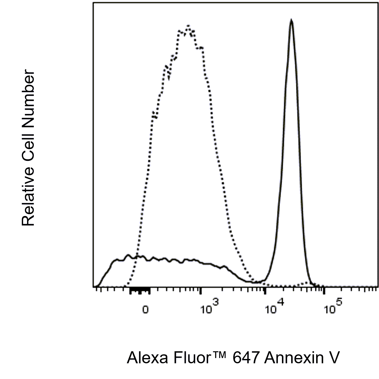
Apoptosis is a normal physiologic process that occurs during embryonic development as well as in maintenance of tissue homeostasis. The apoptotic program is characterized by certain morphologic features, including loss of plasma membrane asymmetry and attachment, condensation of the cytoplasm and nucleus, and internucleosomal cleavage of DNA. Loss of plasma membrane asymmetry is one of the earliest features. In apoptotic cells, the membrane phospholipid phosphatidylserine (PS) is translocated from the inner to the outer leaflet of the plasma membrane, thereby exposing PS to the external cellular environment. Annexin V is a 35-36 kDa Ca2+ dependent phospholipid-binding protein that has a high affinity for PS, and binds to cells with exposed PS. Annexin V may be conjugated to fluorochromes including Alexa Fluor™ 647. This format retains its high affinity for PS and thus serves as a sensitive probe for flow cytometric analysis of cells that are undergoing apoptosis. Since externalization of PS occurs in the earlier stages of apoptosis, Alexa Fluor™ 647 Annexin V staining can identify cells undergoing apoptosis at an earlier stage rather than assays based on nuclear changes such as DNA fragmentation.
Alexa Fluor™ 647 Annexin V staining precedes the loss of membrane integrity which accompanies the latest stages of cell death resulting from either apoptotic or necrotic processes. Therefore, staining withAlexa Fluor™ 647 Annexin V is typically used in conjunction with a vital dye such as 7-Amino-Actinomycin (7-AAD) to allow the investigator to identify early apoptotic cells (Alexa Fluor™ 647 Annexin V positive, 7-AAD negative). Viable cells with intact membranes exclude 7-AAD, whereas the membranes of dead and damaged cells are permeable to the nucleic acid dye, 7-AAD. For example, cells that are considered viable are both Alexa Fluor™ 647 Annexin V negative and 7-AAD negative while cells that are in early apoptosis are Alexa Fluor™ 647 Annexin V positive and 7-AAD negative. Cells that are in late apoptosis or already dead are both Alexa Fluor™ 647 Annexin V positive and 7-AAD positive. This assay does not distinguish between cells that have undergone apoptotic death versus those that have died as a result of a necrotic pathway because in either case, the dead cells will stain with bothAlexa Fluor™ 647 Annexin V and 7-AAD. However, when apoptosis is measured over time, cells can be often tracked from being Alexa Fluor™ 647 Annexin V negative and 7-AAD negative (viable, or no measurable apoptosis), to Alexa Fluor™ 647 Annexin V positive and 7-AAD negative (early apoptosis, membrane integrity is present), and finally to Alexa Fluor™ 647 Annexin V positive and 7-AAD positive (end stage apoptosis and death). The movement of cells through these three stages suggests apoptosis. In contrast, a single observation indicating that cells are both Alexa Fluor™ 647 Annexin V and 7-AAD positive, by itself reveals less information about the process by which the cells underwent their demise.


 下载产品说明书
 用小程序,查商品更便捷


 收藏
收藏
 对比
对比 咨询
咨询
 危险品化学品经营许可证(不带存储) 许可证编号:沪(杨)应急管危经许[2022]202944(QY)
  营业执照(三证合一)