Kit Contents
Description of Components
Material No.
Size
Storage
Vials
Kit Part A
PerCP-Cy5.5 Mouse Anti-BrdU
51-9007682
50 test
4ºC
1
Alexa Fluor™ 647 Mouse Anti-H2AX (pS139)
51-9007683
50 test
4ºC
1
PE Mouse Anti-Cleaved PARP (Asp214) Antibody
51-9007684
50 test
4°C
1
BD Cytofix/Cytoperm™ Fixation/Permeabilization Solution
51-2090KE
25 ml
4°C
1
BD Perm/Wash™ Buffer (10X)
51-2091KE
25 ml
4°C
2
BD Cytofix/Cytoperm™ Plus Permeabilization Buffer
51-2356KC
10 ml
4°C
1
DAPI
51-9007681
100 μl
4°C
1
Kit Part B
BrdU (10 mg/ml)
51-2420KC
5 mg
-80°C
5
DNase
51-2358KC
300 μl
-80°C
5
Apoptosis, DNA Damage and Cell Proliferation Kit
Multiparameter flow cytometry provides a powerful tool for resolving mechanisms by which individual cells in homogenous or mixed cell populations maintain viability, enter and progress through cell cycle or undergo cell death. For this purpose, the Apoptosis, DNA Damage, and Cell Proliferation Kit was designed with the inclusion of fluorescent antibodies specific for incorporated BrdU, phosphorylated H2AX (γH2AX) and cleaved PARP. These probes along with optimized protocols enable multicolor flow cytometric analysis of proliferation, DNA damage and apoptosis, respectively, by individual cells within samples.
Immunofluorescent staining of cells that have incorporated Bromodeoxyuridine (BrdU, an analog of the DNA precursor thymidine) and flow cytometric analysis provides a high resolution technique to determine the frequency and nature of individual cells that have synthesized DNA. Exposure of cells to BrdU in either
in vitro
or
in vivo
experimental model systems allows for BrdU incorporation by actively cycling cell fractions. Pulse labeling of cells with BrdU at various time points, permits the determination of cell-cycle kinetics. Flow cytometric analysis of BrdU incorporation can readily be combined with the simultaneous analysis of cellular phosphorylated H2AX and cleaved PARP levels. Phosphorylated H2AX functions to recruit and localize DNA repair proteins or cell cycle checkpoint factors to DNA-damaged sites. In this way, phosphorylated H2AX promotes DNA repair and maintains genomic stability. Double-stranded DNA breaks caused by replication errors, apoptosis, or other physiological processes (including, immunoglobulin and TCR gene recombinations) and DNA damage caused by ionizing radiation, UV light, or cytotoxic agents lead to H2AX phosphorylation on serine 139, H2AX (pS139), to induce its function. PARP (Poly [ADP-Ribose] Polymerase) is a nuclear chromatin-associated enzyme that is involved in DNA repair. During apoptosis, Caspase-3 cleaves PARP resulting in its inactivation and the inability of cells to repair DNA damage. For this reason, the 89 kDa-cleaved fragment of PARP serves as a marker of cellular apoptosis.
克隆号
(RUO)
组合货号
51-9007685AK/(2 to 8C)+51-9007685BK/(max.-70C)
BD化合物表
描述
数量/尺寸
零件号
EntrezGene ID
Apoptosis, DNA Damage and Cell Proliferation Kit - Part A
N/A
51-9007685AK
N/A
Apoptosis, DNA Damage and Cell Proliferation Kit - Part B
N/A
51-9007685BK
N/A
应用
实验应用
Flow cytometry, Intracellular staining (flow cytometry) (Tested During Development)
目标/特异性
BrdU, H2AX (pS139), Cleaved PARP (Asp214)
参考图片
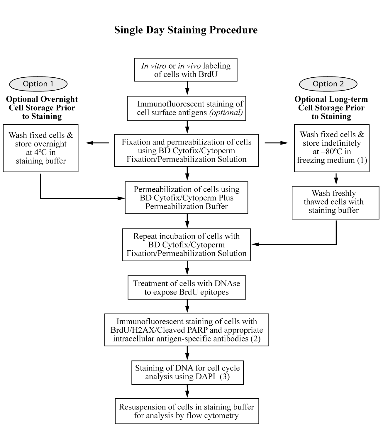
Figure 1. Overview: The BD Pharmingen™ Apoptosis, DNA Damage, and Cell Proliferation Kit Staining Protocol. (1) Recipe for Freezing Media: 10% dimethyl sulfoxide (DMSO) + 90% heat-inactivated Fetal Bovine Serum (FBS). (2) The immunofluorescent staining of cell surface antigens can be done at the same time as staining intracellular antigens provided that the antibodies recognize paraformaldehyde-fixed epitopes. (3) If staining for total DNA content is not desired, then the DAPI staining step can be omitted and fluorescent data for another parameter can then be measured in the UV or Violet channel.
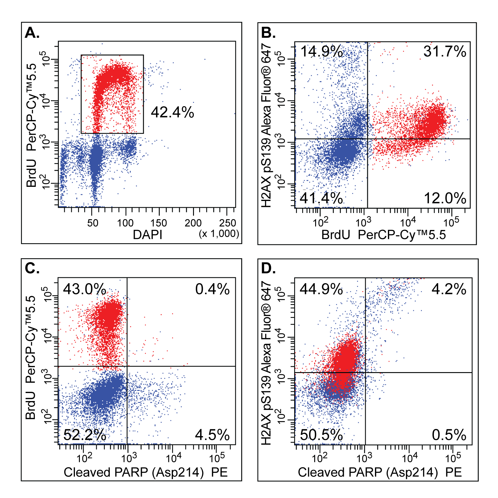
Figure 2. Multiparameter cell cycle analysis of stimulated human peripheral blood mononuclear cells (PBMC). PBMC were stimulated with Purified NA/LE Mouse Anti-Human CD3 (Cat. No. 555329) and Purified NA/LE Mouse Anti-Human CD28 (Cat. No. 555725) antibodies for 3 days. The activated cells were harvested and re-plated in complete medium and labeled with 50 μM BrdU for 1 hr. The cells were then harvested and analyzed by immunofluorescent staining and multicolor flow cytometric analysis using a BD™ LSR II Flow Cytometer System. Panel A: DAPI versus BrdU PerCP-Cy™5.5 staining profile for activated PBMC. Panel B: BrdU PerCP-Cy™5.5 versus H2AX (pS139) Alexa Fluor® 647 profile. Panel C: Cleaved PARP (Asp214) PE versus BrdU PerCP-Cy™5.5 profile. Panel D: Cleaved PARP (Asp214) PE versus H2AX (pS139) Alexa Fluor® 647 profile. BrdU-positive cells are color-gated red whereas BrdU-negative cells are colored blue.


 全部商品分类


 下载产品说明书
 用小程序,查商品更便捷


 收藏
收藏
 对比
对比 咨询
咨询