


1/27

品牌: CST
 下载产品说明书
 用小程序,查商品更便捷


 收藏
收藏
 对比
对比 咨询
咨询
产品介绍
产品信息
抗原名称
ApoptosisNecroptosis

来源纯化
Monoclonal antibodies are produced by immunizing animals with synthetic peptides surrounding Leu190 of human RIP1, the carboxy terminus of human MLKL and human caspase-8, the amino-terminal sequence of p10 of human caspase-8, amino terminal residues adjacent to Asp175 of human caspase-3, a recombinant protein corresponding to the p20 subunit of caspase-3, and phosphopeptides surrounding human Ser166 of human RIP and Ser358 of human MLKL.
简单描述
Antibody Sampler Kit for studying RIPK1/RIPK1 (Ser166) phosphate/Casp8 (Asp384) cleaved/Casp8/MLKL/MLKL (Thr357/Ser358) phosphate/Casp3/Casp3 (Asp175) cleaved in the research area.
研究领域
癌症,细胞生物学,纤维化,神经科学,
应用
目标/特异性
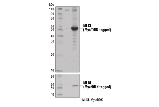
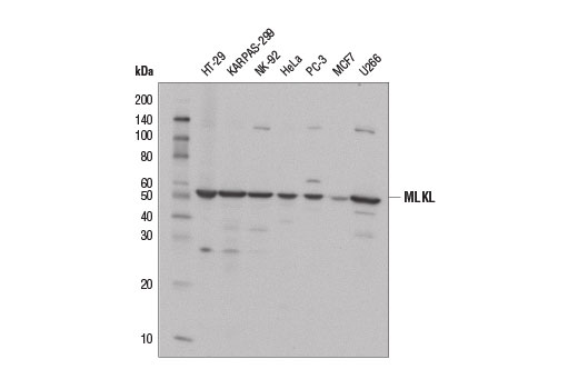
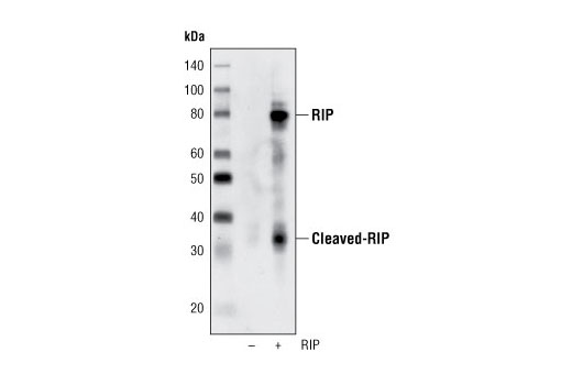
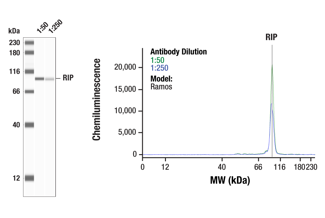
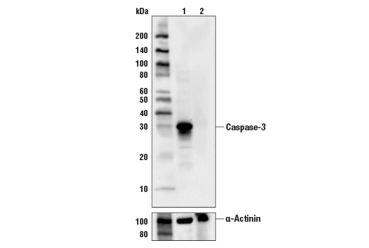
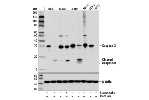
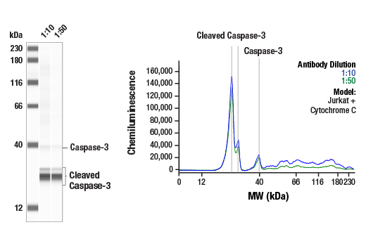
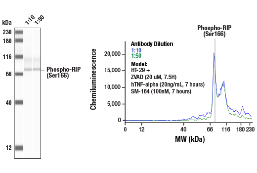
Specificity/Sensitivity
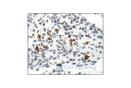
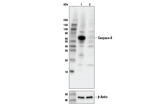
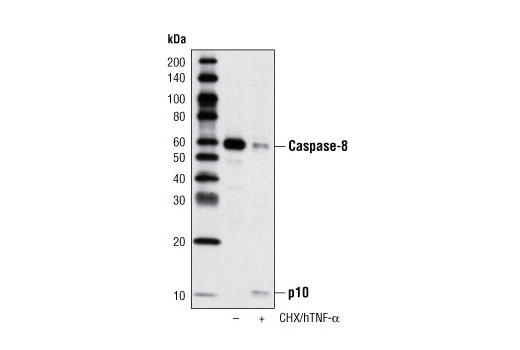
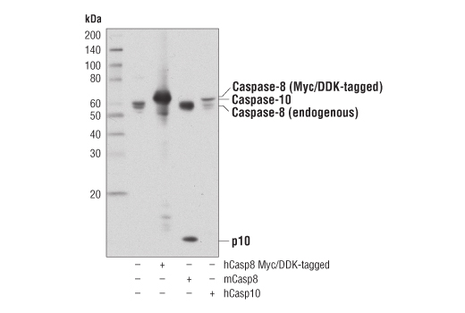
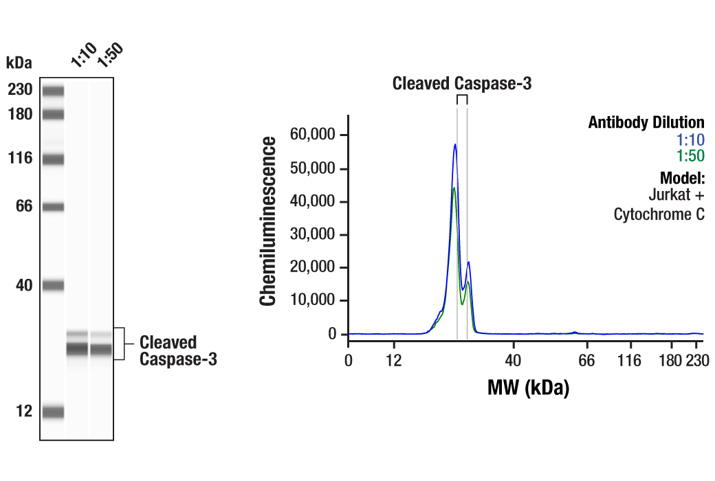
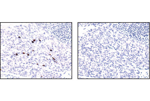
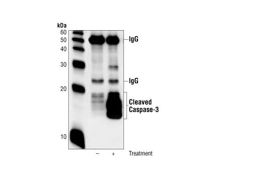
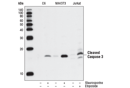
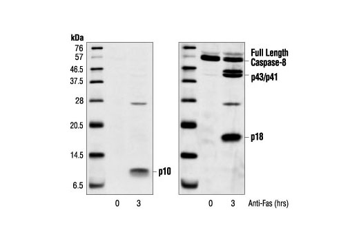
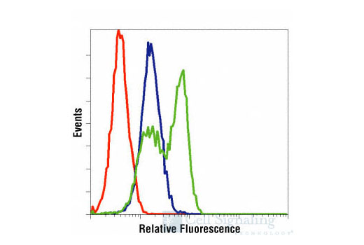
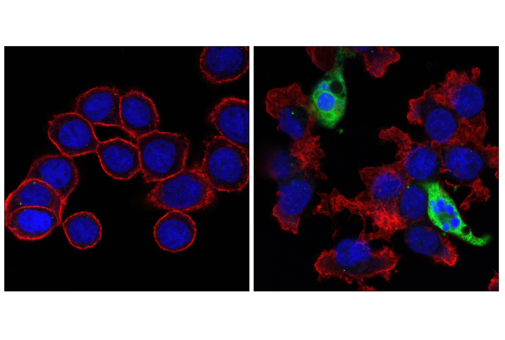
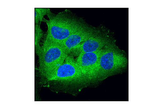
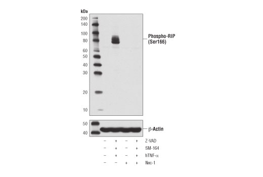
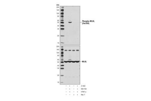
Each antibody in the Apoptosis/Necroptosis Antibody Sampler Kit detects endogenous levels of its target protein. MLKL (D2I6N) Rabbit mAb cross-reacts with a band at 130 kDa in some cell lines. Phospho-MLKL (Ser358) (D6H3V) Rabbit mAb may also react with MLKL when dually phoshorylated at Thr357 and Ser358. Caspase-3 (D3R6Y) Rabbit mAb detects full-length caspase-3 as well as the large subunit (p20) of caspase-3 resulting from cleavage during apoptosis. Cleaved Caspase-3 (Asp175) (5A1) Rabbit mAb detects endogenous levels of the large subunit of caspase-3 but does not detect full-length caspase-3 or other cleaved caspases. Cleaved Caspase-8 (Asp384) (11G10) Mouse mAb detects endogenous levels of the small fragment of caspase-8 resulting from cleavage at aspartic acid 384. The antibody does not cross-react with full length caspase-8. Caspase-8 (D35G2) Rabbit mAb detects endogenous levels of total caspase-8, including the p10 subunit of the activated protein. It may also cross-react with overexpressed levels of caspase-10.
背景
背景
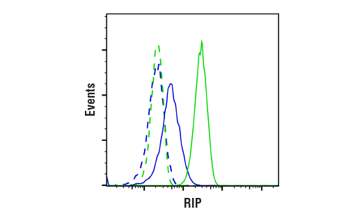
Apoptosis is a regulated physiological process leading to cell death (1,2). Caspases, a family of cysteine acid proteases, are central regulators of apoptosis. Caspases are synthesized as inactive zymogens containing a pro-domain followed by large (p20) and small subunits (p10) that are proteolytically processed in a cascade of caspase activity. Initiator caspases (including 8, 9, 10, and 12) are closely coupled to proapoptotic signals. Once activated, these caspases cleave and activate downstream effector caspases (including 3, 6, and 7), which in turn cleave cytoskeletal and nuclear proteins like PARP, α-fodrin, DFF, and lamin A, and induce apoptosis. Cytochrome c released from mitochondria is coupled to the activation of caspase-9, a key initiator caspase. Apoptosis induced through the extrinsic mechanisms involving death receptors in the tumor necrosis factor receptor superfamily activates caspase-8. Activated caspase-8 cleaves and activates downstream effector caspases, such as caspase-1, -3, -6, and -7. Caspase-3 is a critical executioner of apoptosis, as it is either partially or totally responsible for the proteolytic cleavage of many key proteins, such as the nuclear enzyme poly (ADP-ribose) polymerase (PARP).Necroptosis, a regulated pathway for necrotic cell death, is triggered by a number of inflammatory signals, including cytokines in the tumor necrosis factor (TNF) family, pathogen sensors such as toll-like receptors (TLRs), and ischemic injury (3,4). Necroptosis is negatively regulated by caspase-8 mediated apoptosis in which the kinase RIP/RIPK1 is cleaved (5). Furthermore, necroptosis is inhibited by a small molecule inhibitor of RIP, necrostatin-1 (Nec-1) (6). Research studies show that necroptosis contributes to a number of pathological conditions, and Nec-1 has been shown to provide neuroprotection in models such as ischemic brain injury (7). RIP is phosphorylated at several sites within the kinase domain that are sensitive to Nec-1, including Ser14, Ser15, Ser161, and Ser166 (8). Phosphorylation drives association with RIP3, which is required for necroptosis (9-11). Mixed lineage kinase domain-like protein (MLKL) is a pseudokinase that was identified as a downstream target of RIP3 in the necroptosis pathway (12). During necroptosis, RIP3 is phosphorylated at Ser227, which recruits MLKL and leads to its phosphorylation at Thr357 and Ser358 (12). Knockdown of MLKL through multiple mechanisms results in inhibition of necroptosis (13). Phosphorylation of MLKL during necroptosis leads to its oligomerization with pore formation that affects membrane integrity (14-17).
1.Degterev, A. et al. (2003) Oncogene 22, 8543-67.
2.Green, D.R. (1998) Cell 94, 695-8.
3.Christofferson, D.E. and Yuan, J. (2010) Curr Opin Cell Biol 22, 263-8.
4.Kaczmarek, A. et al. (2013) Immunity 38, 209-23.
5.Lin, Y. et al. (1999) Genes Dev 13, 2514-26.
6.Degterev, A. et al. (2008) Nat Chem Biol 4, 313-21.
7.Degterev, A. et al. (2005) Nat Chem Biol 1, 112-9.
8.Ofengeim, D. and Yuan, J. (2013) Nat Rev Mol Cell Biol 14, 727-36.
9.Cho, Y.S. et al. (2009) Cell 137, 1112-23.
10.He, S. et al. (2009) Cell 137, 1100-11.
11.Zhang, D.W. et al. (2009) Science 325, 332-6.
12.Sun, L. et al. (2012) Cell 148, 213-27.
13.Wu, J. et al. (2013) Cell Res 23, 994-1006.
14.Cai, Z. et al. (2014) Nat Cell Biol 16, 55-65.
15.Chen, X. et al. (2014) Cell Res 24, 105-21.
16.Wang, H. et al. (2014) Mol Cell 54, 133-46.
17.Dondelinger, Y. et al. (2014) Cell Rep 7, 971-81.
研究领域
癌症,细胞生物学,纤维化,神经科学,
数据库链接
Entrez-Gene ID
8737,841,197259,836
UniProt ID
Q13546,Q14790,Q8NB16,P42574
参考图片
声明 :本官网所有报价均为常温或者蓝冰运输价格,如有产品需要干冰运输,需另外加收干冰运输费。

 危险品化学品经营许可证(不带存储) 许可证编号:沪(杨)应急管危经许[2022]202944(QY)
  营业执照(三证合一)