


 下载产品说明书
 用小程序,查商品更便捷


 收藏
收藏
 对比
对比 咨询
咨询
Specificity/Sensitivity
参考图片
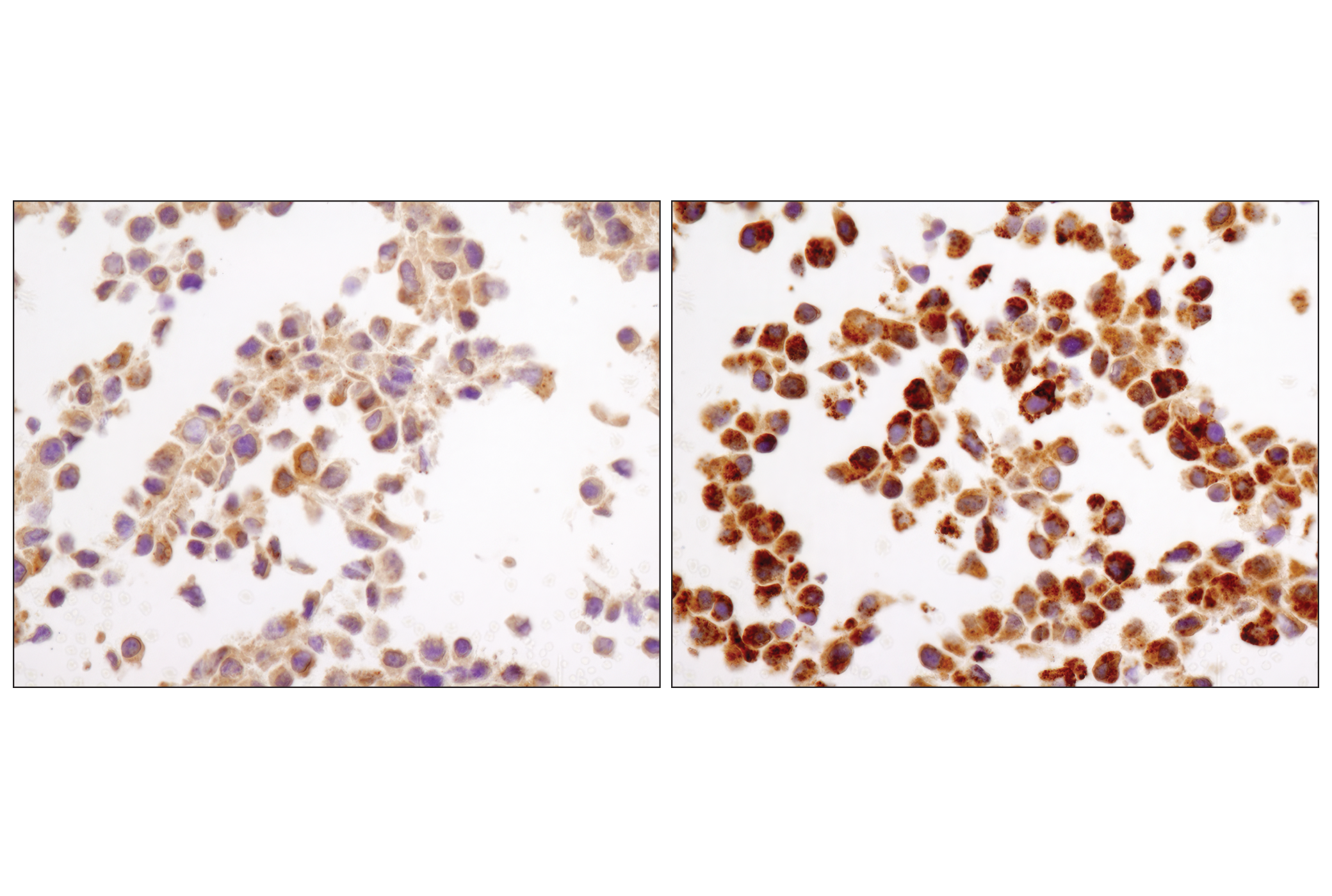
Immunohistochemical analysis of paraffin-embedded NIH/3T3 cell pellets, control (left) or chloroquine-treated (right), using LC3A/B (D3U4C) XP® Rabbit mAb.
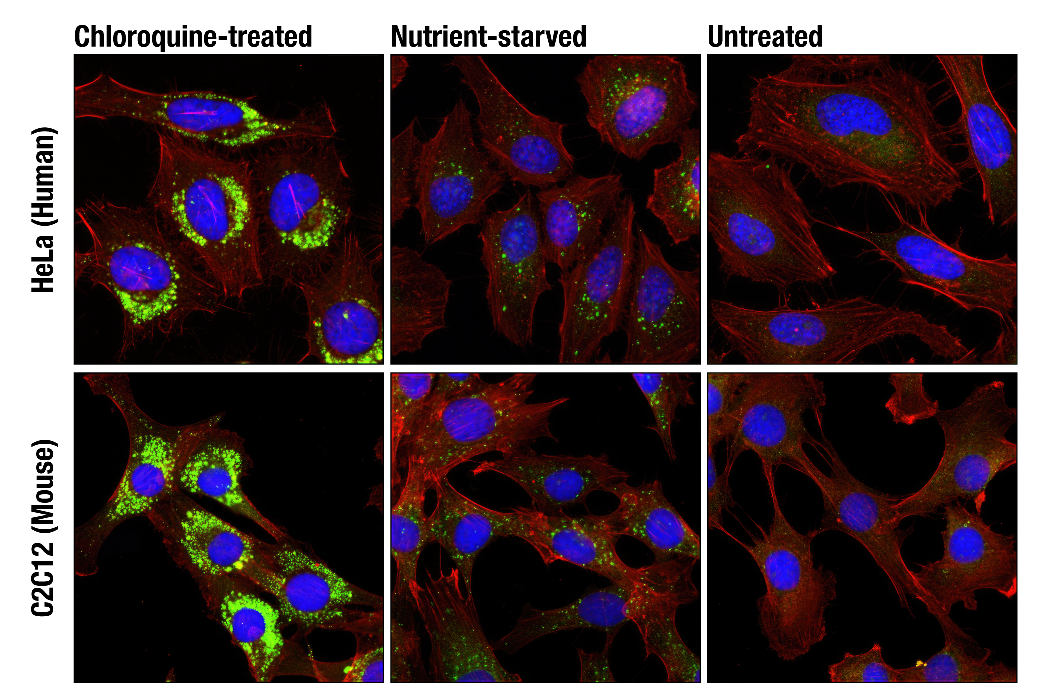
Confocal immunofluorescent analysis of HeLa (upper) and C2C12 (lower) cells, chloroquine-treated (50 μM, overnight; left), nutrient-starved with EBSS (3 hr, middle) or untreated (right) using LC3A/B (D3U4C) XP® Rabbit mAb (green) and β-Actin (13E5) Rabbit mAb (Alexa Fluor® 555 Conjugate) #8046 (red). Blue pseudocolor= DRAQ5® #4084 (fluorescent DNA dye).
After the primary antibody is bound to the target protein, a complex with HRP-linked secondary antibody is formed. The LumiGLO* is added and emits light during enzyme catalyzed decomposition.
Western blot analysis of extracts from various cell lines using Atg3 Antibody.
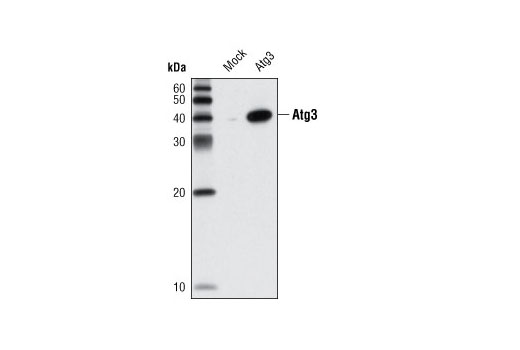
Western blot analysis of extracts from HeLa cells, mock transfected or transfected with mouse Atg3, using Atg3 Antibody.
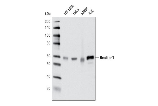
Western blot analysis of extracts from various cell lines using Beclin-1 (D40C5) Rabbit mAb.
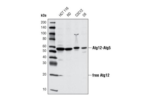
Western blot analysis of extracts from various cell lines using Atg12 (D88H11) Rabbit mAb.
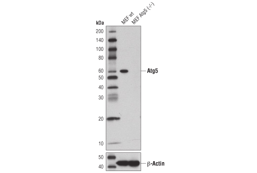
Western blot analysis of extracts from wild-type MEFs (wt) or MEFs from Atg5 knockouts (Atg5-/-) using Atg5 (D5F5U) Rabbit mAb (upper), or β-Actin (D6A8) Rabbit mAb #8457 (lower). Atg5-/- MEFs were kindly provided by Dr. Ramnik Xavier, Massachusetts General Hospital, Harvard Medical School, Boston, MA.
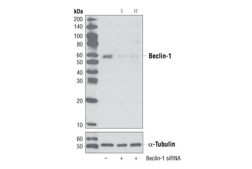
Western blot analysis of extracts from HeLa cells, transfected with 100 nM SignalSilence® Control siRNA (Unconjugated) #6568 (-), SignalSilence® Beclin-1 siRNA I #6222 (+) or SignalSilence® Beclin-1 siRNA II (+), using Beclin-1 (D40C5) XP® Rabbit mAb #3495 (upper) or α-Tubulin (11H10) Rabbit mAb #2125 (lower). The Beclin-1 (D40C5) XP® Rabbit mAb confirms silencing of Beclin-1 expression, while the α-Tubulin (11H10) Rabbit mAb is used to control for loading and specificity of Beclin-1 siRNA.
Western blot analysis of extracts from various cell lines using Atg12 (D88H11) Rabbit mAb #4180.
Western blot 分析多种细胞系的细胞提取物,使用抗体为Atg12 (D88H11) Rabbit mAb 兔单抗#4180。
Western blot analysis of extracts from various cell lines using Beclin-1 (D40C5) Rabbit mAb #3495.
Western blot 分析多种细胞系的细胞提取物,使用抗体是Beclin-1 (D40C5) Rabbit mAb 兔单抗#3495。
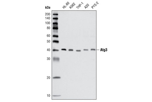
Western blot analysis of extracts from various cell lines using Atg3 Antibody #3415.
Western blot分析多种细胞系的细胞提取物,使用抗体是Atg3 Antibody #3415。
Immunoprecipitation of Atg5 from HT-1080 cells using Atg5 (D1G9) Rabbit mAb #8540. Western blot detection was performed using the same antibody (upper), or with Atg12 (D88H11) Rabbit mAb #4180 (lower). Lane 1 is 10% input, lane 2 is precipitated with non-specific rabbit IgG, lane 3 is precipitated with Atg5 (D1G9) Rabbit mAb #8540.
使用Atg5 (D1G9) Rabbit mAb #8540对HT-1080细胞进行免疫共沉淀分析。Western blot 检测使用相同的抗体(上图),Atg12 (D88H11) Rabbit mAb 兔单抗#4180(下图)。泳道1是10% input,泳道2为非特异性lgG沉淀的样品,泳道3为Atg5 (D1G9) Rabbit mAb 兔单抗#8540沉淀的样品。
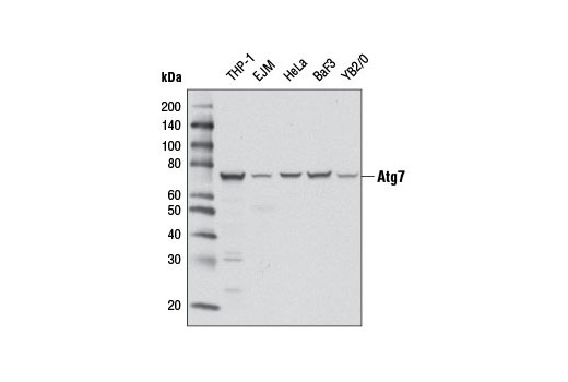
Western blot analysis of extracts from various cell lines using Atg7 (D12B11) Rabbit mAb.
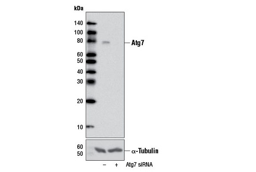
Western blot analysis of extracts from HeLa cells, transfected with 100 nM SignalSilence® Control siRNA (Unconjugated) #6568 (-) or SignalSilence® Atg7 siRNA I #6604 (+), using Atg7 (D12B11) Rabbit mAb (upper) or α-Tubulin (11Η10) Rabbit mAb #2125 (lower). The Atg7 (D12B11) Rabbit mAb confirms silencing of Atg7 expression, while the α-Tubulin (11H10) Rabbit mAb is used as a loading control.
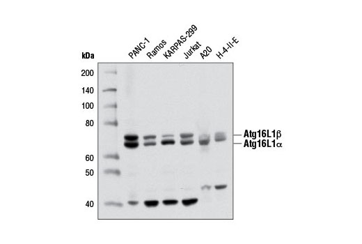
Western blot analysis of extracts from various cell lines using Atg16L1 (D6D5) Rabbit mAb.
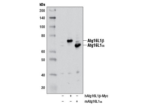
Western blot analysis of extracts from 293T cells, mock transfected (-) or transfected with either a myc-tagged human Atg16L1β construct (hAtg16L1β-Myc; +) or a mouse Atg16L1α construct (mAtg16L1α; +), using Atg16L1 (D6D5) Rabbit mAb. The myc-tagged human Atg16L1β construct was kindly provided by Dr. Qing Zhong, University of California Berkeley.
Confocal immunofluorescent analysis of EBSS-starved PANC-1 cells using Atg16L1 (D6D5) Rabbit mAb (green). Blue pseudocolor = DRAQ5® #4084 (fluorescent DNA dye).
Western blot analysis of extracts from various cell lines using Atg7 (D12B11) Rabbit mAb #8558.
Western blot分析多种细胞系的细胞提取物,使用抗体是Atg7 (D12B11) Rabbit mAb 兔单抗#8558。
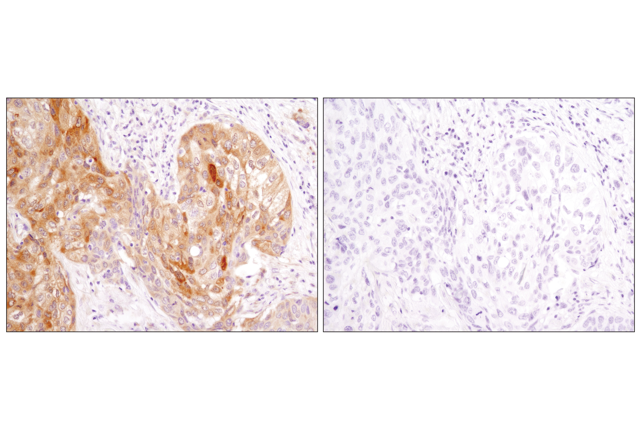
Immunohistochemical analysis of paraffin-embedded human lung carcinoma using LC3A/B (D3U4C) XP® Rabbit mAb in the presence of control peptide (left) or antigen-specific peptide (right).
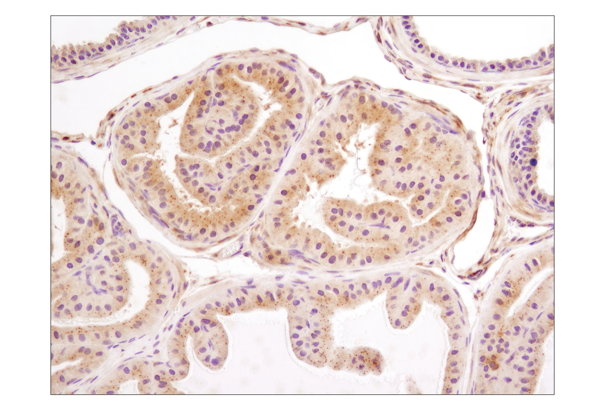
Immunohistochemical analysis of paraffin-embedded mouse prostate using LC3A/B (D3U4C) XP® Rabbit mAb.
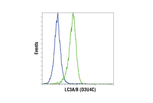
Flow cytometric analysis of HeLa cells, untreated (blue) or treated with chloroquine (50 µM, 16 hr) (green), using LC3A/B (D3U4C) Rabbit mAb. Anti-rabbit IgG (H+L), F(ab')2 Fragment (Alexa Fluor® 647 Conjugate) #4414 was used as a secondary antibody.
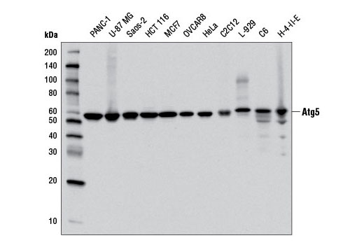
Western blot analysis of extracts from various cell lines using Atg5 (D5F5U) Rabbit mAb.
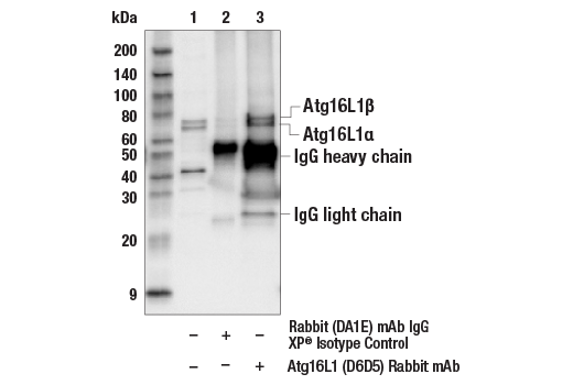
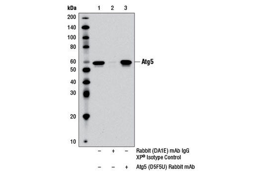
Immunoprecipitation of Atg5 from PANC-1 cell extracts using Rabbit (DA1E) mAb IgG XP® Isotype Control #3900 (lane 2) or Atg5 (D5F5U) Rabbit mAb (lane 3). Lane 1 is 10% input. Western blot was performed using Atg5 (D5F5U) Rabbit mAb. Mouse Anti-rabbit IgG (Conformation Specific) (L27A9) mAb #3678 was used as a secondary antibody to avoid cross-reactivity with rabbit IgG.
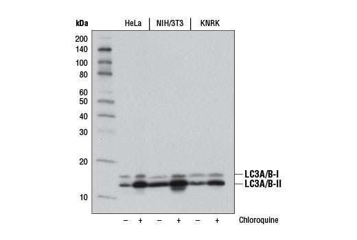
Western blot analysis of extracts from HeLa, NIH/3T3, and KNRK cells, untreated (-) or chloroquine-treated (50 μM, overnight; +), using LC3A/B (D3U4C) XP® Rabbit mAb #12741.
Western blot analysis of extracts from various cell lines using Atg5 (D5F5U) Rabbit mAb #12994.
Western blot analysis of extracts from various cell lines using Atg16L1 (D6D5) Rabbit mAb #8089.
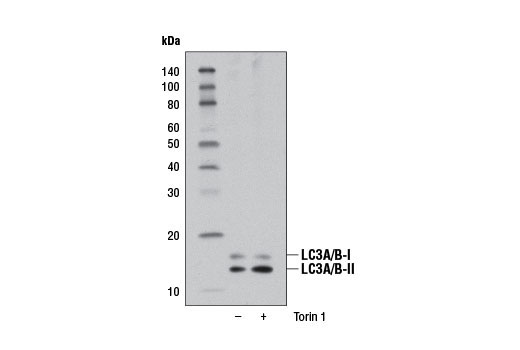
Western blot analysis of extracts from RD cells, untreated (-) or Torin 1-treated (250 nM, 4 hr; +), using LC3A/B (D3U4C) XP® Rabbit mAb.
Western blot analysis of extracts from HeLa, NIH/3T3, and KNRK cells, untreated (-) or chloroquine-treated (50 μM, overnight; +), using LC3A/B (D3U4C) XP® Rabbit mAb.

 危险品化学品经营许可证(不带存储) 许可证编号:沪(杨)应急管危经许[2022]202944(QY)
  营业执照(三证合一)