


 下载产品说明书 下载SDS
 用小程序,查商品更便捷


 收藏
收藏
 对比
对比 咨询
咨询
- 描述EntrezGene ID
- Biotin Mouse B Lymphocyte Enrichment CocktailN/A
- Streptavidin Particles Plus - DMN/A
参考图片
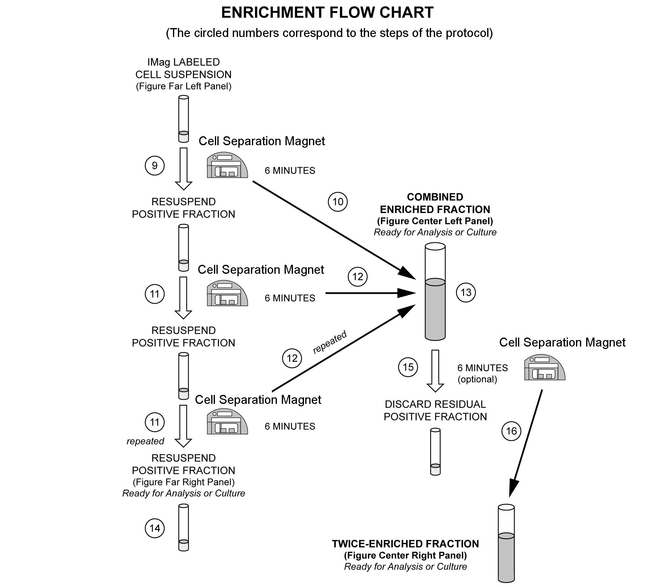
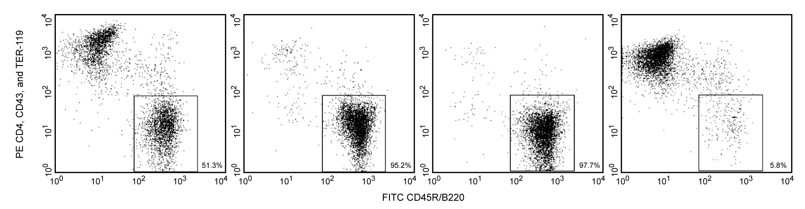
Enrichment of B lymphocytes from mouse spleen. BALB/c splenocytes were labeled with the BD IMag™ Mouse B Lymphocyte Enrichment Set - DM (Cat. No. 557792) and separated on the BD IMag™ Cell Separation Magnet (Cat. No. 552311) according to the accompanying protocol. To demonstrate the efficiency of the enrichment, cells were stained with FITC Anti-Mouse CD45R/B220 (Cat. No. 553087/553088) to detect B lymphocytes and a mixture of PE Rat Anti-Mouse CD4 (Cat. No. 557308/553730), CD43(Cat. No. 553271), and TER119/Erythroid Cells (Cat. No. 553673) monoclonal antibodies to detect non-B leukocytes and erythrocytes. Dead cells were excluded by staining with Propidium Iodide Staining Solution (Cat. No. 556463). Flow cytometry was performed on a BD FACSCalibur™ flow cytometry system. Please refer to the Enrichment Flow Chart to identify the cell populations represented in this figure. The percentage of B cells is indicated in the lower-right corner of each panel. The far left panel shows unseparated splenocytes. The middle left panel shows the combined enriched fraction after three 6minute magnetic separations. The middle right panel shows the twice-enriched fraction after an additional 6-minute separation of the cells shown in the middle left panel. This additional incubation can result in up to 5% increased purity with up to a 5% decrease in recovery. The far right panel shows the positive fraction.
Enrichment of B lymphocytes from mouse spleen. BALB/c splenocytes were labeled with the BD IMag™ Mouse B Lymphocyte Enrichment Set - DM (Cat. No. 557792) and separated on the BD IMag™ Cell Separation Magnet (Cat. No. 552311) according to the accompanying protocol. To demonstrate the efficiency of the enrichment, cells were stained with FITC Anti-Mouse CD45R/B220 (Cat. No. 553087/553088) to detect B lymphocytes and a mixture of PE Rat Anti-Mouse CD4 (Cat. No. 557308/553730), CD43(Cat. No. 553271), and TER119/Erythroid Cells (Cat. No. 553673) monoclonal antibodies to detect non-B leukocytes and erythrocytes. Dead cells were excluded by staining with Propidium Iodide Staining Solution (Cat. No. 556463). Flow cytometry was performed on a BD FACSCalibur™ flow cytometry system. Please refer to the Enrichment Flow Chart to identify the cell populations represented in this figure. The percentage of B cells is indicated in the lower-right corner of each panel. The far left panel shows unseparated splenocytes. The middle left panel shows the combined enriched fraction after three 6minute magnetic separations. The middle right panel shows the twice-enriched fraction after an additional 6-minute separation of the cells shown in the middle left panel. This additional incubation can result in up to 5% increased purity with up to a 5% decrease in recovery. The far right panel shows the positive fraction.

 危险品化学品经营许可证(不带存储) 许可证编号:沪(杨)应急管危经许[2022]202944(QY)
  营业执照(三证合一)