


 下载产品说明书
 用小程序,查商品更便捷


 收藏
收藏
 对比
对比 咨询
咨询
- 描述数量/尺寸零件号EntrezGene ID
- APO-BRDU™ Kit Part AN/A51-6576AKN/A
- APO-BRDU™ Kit Part BN/A51-6576BKN/A
参考图片
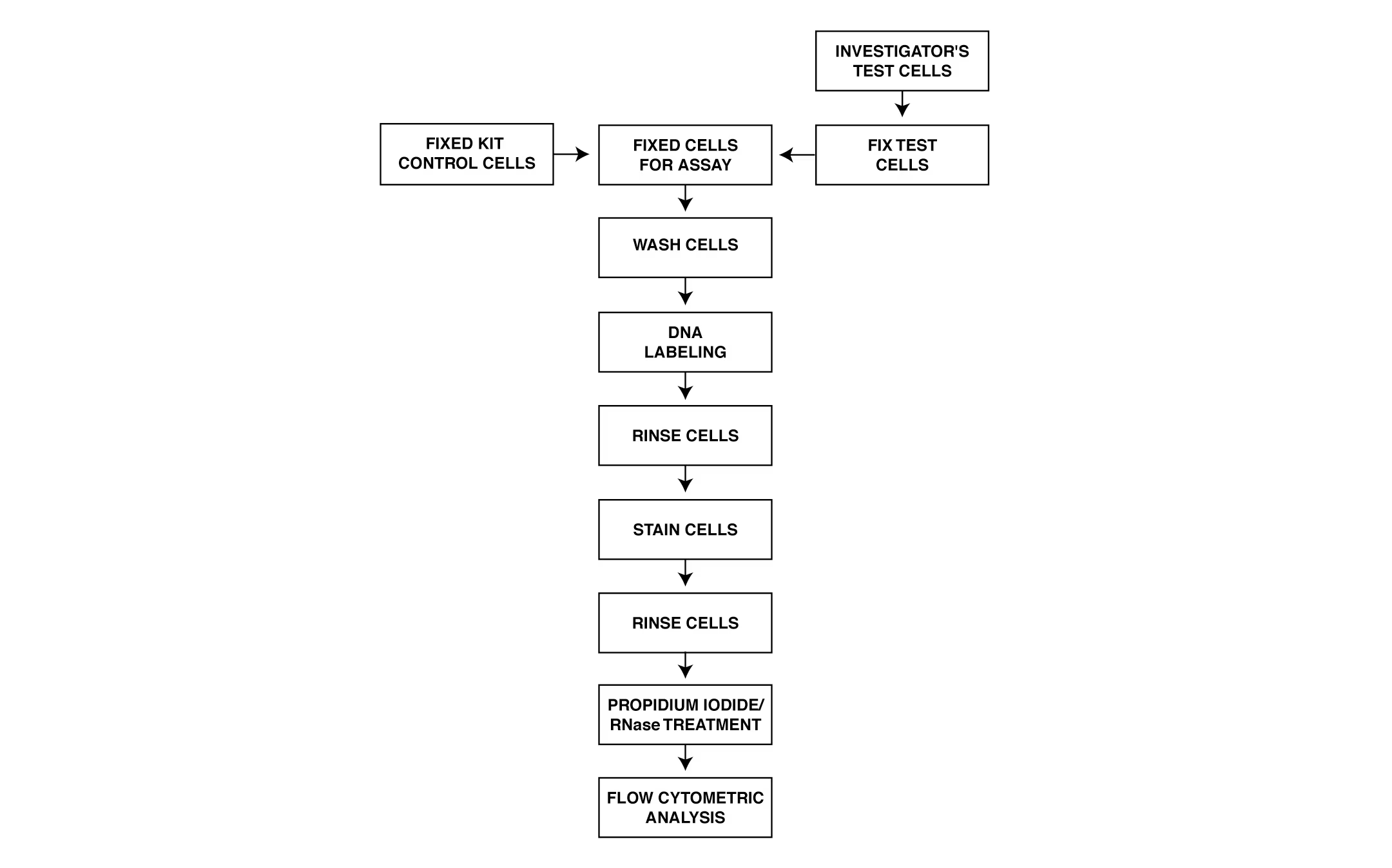
Schematic Representation of APO-BRDU™ Labeling. The enzyme deoxynucleotidyl transferase (TdT) catalyzes a template dependent addition of bromolated deoxyuridine triphosphates (Br-dUTP) to the 3'-hydroxyl ends of double- and single-stranded DNA. After Br-dUTP incorporation, DNA break sites are identified by a FITC-labeled anti-BrdU mAb.
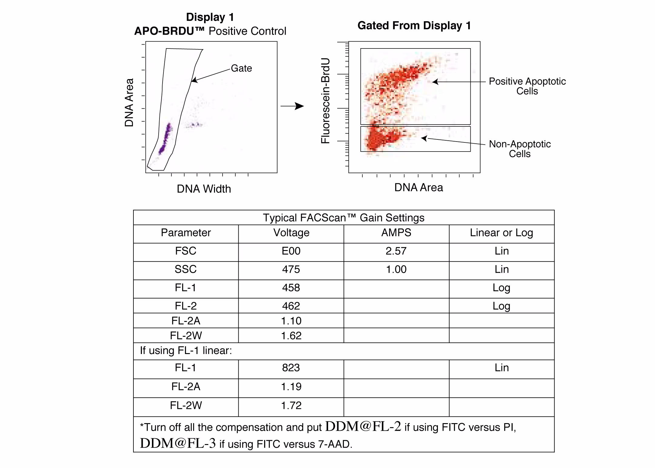
Flow Cytometer Setup for Becton Dickinson Hardware. Positive control cells were labeled with both PI (DNA) and FITC-BrdU mAb. Display 1: Non-clumped cells are gated. Gated From Display 1: Separate boxes are drawn around cells that stain positive (upper box) and negative (lower box) with the FITC-BrdU mAb. An example of FACScan™ gain settings is shown.
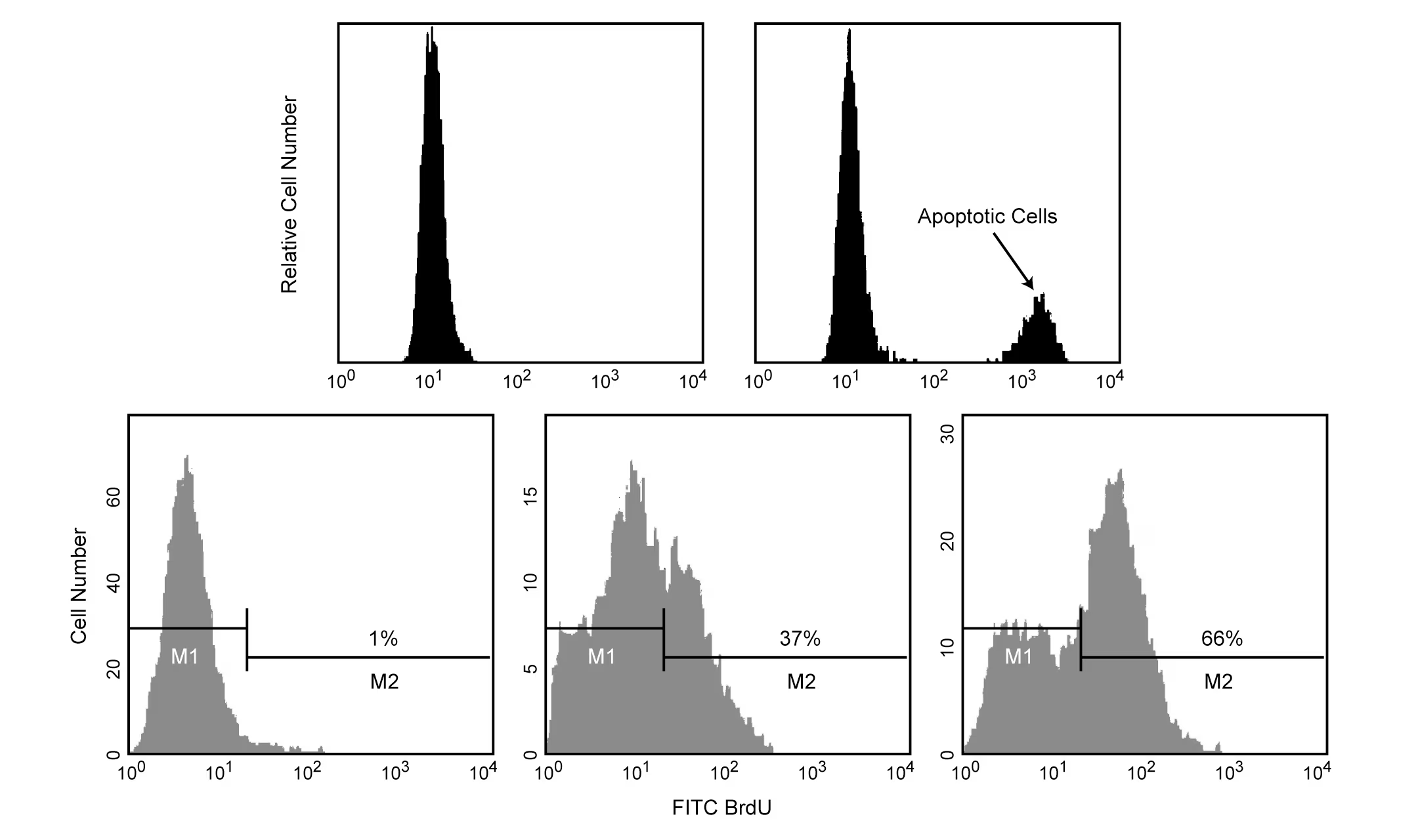
Flow Cytometry Data of APO-BRDU™ Negative and Positive Control cells (top row). Negative and Positive control cells were incubated with Br-dUTP in the presence of TdT enzyme in order to incorporate Br-dUTP into exposed 3'-OH DNA ends. Br-dUTP sites are detected with a FITC-labeled anti-BrdU mAb. Non-apoptotic cells do not incorporate significant amounts of Br-dUTP due to the lack of exposed 3'-OH ends, and consequently have relatively little fluorescence compared to apoptotic cells which have an abundance of 3'-OH ends. Flow Cytometric Analysis of HPB-ALL Human Leukemia Cells Using APO-BRDU™ (bottom row). HPB-ALL human leukemia cells were left untreated (left panel) or treated with anti-human Fas mAb, clone DX2 (Cat. No. 33450D) and Protein G for 2 hr (middle panel) or 12 hr (right panel). Cells were fixed and incubated with Br-dUTP in the presence of TdT enzyme in order to incorporate Br-dUTP into exposed 3'-OH DNA ends. Br-dUTP was detected with a FITC-labeled anti-BrdU mAb. Non-apoptotic cells (M1 gates) do not incorporate significant amounts of Br-dUTP due to lack of exposed 3'-OH ends, and consequently have relatively little fluorescence compared to apoptotic cells which have an abundance of 3'-OH end (M2 gates). DX2-induced, Fas-mediated apoptosis is shown by increases in the number of cells staining with anti-BrdU-FITC mAb (M2 gates) after 2 and 12 hr. The M1 and M2 gates demarcate non-apoptotic and apoptotic populations, respectively.

 危险品化学品经营许可证(不带存储) 许可证编号:沪(杨)应急管危经许[2022]202944(QY)
  营业执照(三证合一)