


 用小程序,查商品更便捷


Product Usage Information
| Application | Dilution |
|---|---|
| Western Blotting | 1:1000 |
| Immunoprecipitation | 1:50 |
| IHC Leica Bond | 1:25 - 1:100 |
| Immunohistochemistry (Paraffin) | 1:50 - 1:200 |
| Immunofluorescence (Immunocytochemistry) | 1:400 - 1:1600 |
Specificity/Sensitivity
Species Reactivity:
Human, Mouse
参考图片
Confocal immunofluorescent analysis of A172 (positive, left) and MCF7 (negative, right) cells using N-Cadherin (D4R1H) XP® Rabbit mAb (green). Blue pseudocolor= DRAQ5® #4084 (fluorescent DNA dye).使用N-Cadherin (D4R1H) XP® Rabbit mAb(绿色)对A172 (阳性,左)和MCF7 (阴性,右)进行激光共聚焦免疫荧光分析。蓝色假色= DRAQ5®#4084 (fluorescent DNA dye)。
Immunohistochemical analysis of paraffin-embedded human ovarian carcinoma using N-Cadherin (D4R1H) XP® Rabbit mAb.使用N-Cadherin (D4R1H) XP® Rabbit mAb对石蜡包埋的人卵巢癌组织进行免疫组化分析。
Immunohistochemical analysis of paraffin-embedded A172 (positive, left) and MCF7 (negative, right) cell pellets using N-Cadherin (D4R1H) XP® Rabbit mAb.使用N-Cadherin (D4R1H) XP® Rabbit mAb对石蜡包埋的A172 (阳性,左)和MCF7 (阴性,右)进行免疫组化分析。
Immunohistochemical analysis of paraffin-embedded human colon using N-Cadherin (D4R1H) XP® Rabbit mAb. Note staining of myenteric plexus. 使用N-Cadherin (D4R1H) XP®Rabbit mAb对石蜡包埋的人结肠癌组织进行免疫组化分析。注意肌间神经丛的染色。
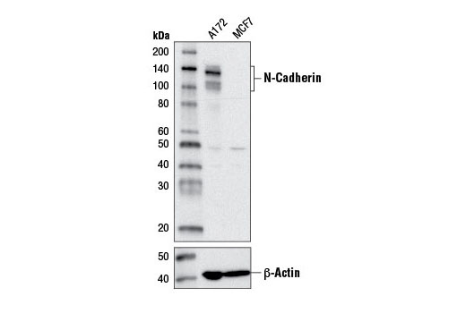
Western blot analysis of extracts from A172 and MCF7 cells using N-Cadherin (D4R1H) XP® Rabbit mAb (upper) or β-Actin (D6A8) Rabbit mAb #8457 (lower).使用N-Cadherin (D4R1H) XP® Rabbit mAb (上) 或β-Actin (D6A8) Rabbit mAb #8457 (下)对A172和MCF7细胞提取物进行western blot分析。

 危险品化学品经营许可证(不带存储) 许可证编号:沪(杨)应急管危经许[2022]202944(QY)
  营业执照(三证合一)