


 下载产品说明书 下载SDS
 用小程序,查商品更便捷


 收藏
收藏
 对比
对比 咨询
咨询

参考图片
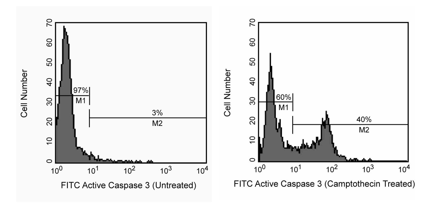
Flow cytometric analysis of apoptotic and non-apoptotic populations for active caspase-3. Jurkat cells (Human T-cell leukemia; ATCC TIB-152) were left untreated (left panel) or treated with 4 µM of camptothecin for 4 hr to induce apoptosis (right panel). Cells were washed once in PBS, then fixed and permeabilized using the BD Cytofix/Cytoperm™ Kit (Cat. No. 554714) for 20 min at room temperature (RT), pelleted and washed with BD Perm/Wash™ buffer (component of Cat. No. 554714). Cells were subsequently stained with the FITC rabbit anti- active caspase-3 antibody (clone C92-605). Cells were then washed and resuspended in BD Perm/Wash™ buffer before analyzing by flow cytometry. The results show that untreated cells were primarily negative for active caspase-3 (left panel, M1); whereas over one third of the treated cells were positive for active caspase-3 staining (right panel, M2).

 危险品化学品经营许可证(不带存储) 许可证编号:沪(杨)应急管危经许[2022]202944(QY)
  营业执照(三证合一)