


 下载产品说明书 下载SDS
 用小程序,查商品更便捷


 收藏
收藏
 对比
对比 咨询
咨询
Met1-Leu352
Accession # P51681
Scientific Data
.") View Larger
View LargerCCR5 in Human Dorsal Root Ganglia. CCR5 was detected in immersion fixed paraffin-embedded sections of human dorsal root ganglia using 25 µg/mL Human CCR5 Monoclonal Antibody (Catalog # MAB181) overnight at 4 °C. Tissue was stained with the Anti-Mouse HRP-AEC Cell & Tissue Staining Kit (red; Catalog # CTS003) and counterstained with hematoxylin (blue). View our protocol for Chromogenic IHC Staining of Paraffin-embedded Tissue Sections.
 View Larger
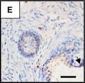
Detection of Human CCR5 by Immunohistochemistry Presence of potential HIV target cells in the human prostate. Immunohistochemistry on uninfected prostate sections before culture showed the presence of periglandular foci of HLA-DR+ (A) and CD4+ cells (B, serial section with A) as well as scattered stromal cells staining positive for HLA-DR (A), CD4 (B), CD3 (C), CD68 (D), CCR5 (E) and CXCR4 (F). The arrows point out immune cells inserted within the epithelium-Scale bars = 50 μm; (G): the respective proportions of CD3, CD4, CD68, CXCR4 and CCR5+ cells per surface unit were evaluated on whole prostate sections from a minimum of 3 donors whose explants were subsequently exposed to HIV-1 strains. Image collected and cropped by CiteAb from the following publication (https://pubmed.ncbi.nlm.nih.gov/19117522), licensed under a CC-BY license. Not internally tested by R&D Systems.
 View Larger
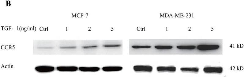
Detection of Human CCR5 by Western Blot TGF-beta signaling regulated the expression of CCR5(A) 3×10⁵ MDA-MB-231 and MCF-7 cells were stimulated with 1-5ng/ml TGF-beta 1 for 24 h, and total RNA was isolated and tested for CCR5 mRNA by quantitative PCR. (B) Western blot for CCR5 protein in breast cancer cells (106) under TGF-beta 1 stimulation for 48 h. Data presented were representatives of at least three independent experiments. (C) MDA-MB-231 and MCF-7 cells (3×10⁵) were co-transfected with pGL3-CCR5 and pRL-TK and exposed to different concentrations of TGF-beta 1 for 24 h, and luciferase activities were determined. (D) MDA-MB-231 and MCF-7 cells were pre-treated with 5μM SIS3 for 2 h, and cells were subjected to luciferase assay. (E) 106 MCF-7 cells were transfected with TGF beta RI/ALK5 siRNA, and were then co-cultured with lactate-activated THP-1 macrophages (ratio 1:1) for 24 h. The protein levels of CCR5 were assayed by western blot. (F) The expression of TGF-beta 1, CCL5 and CCR5 in clinical samples obtained from breast cancer patients. The mRNA levels were measured by quantitative PCR, and the correlation between TGF-beta 1 and CCL5-CCR5 axis was shown. (G) Representative IHC staining for TGF-beta 1, CCL5 and CCR5 in breast cancer samples. The sample used was derived from 28 breast cancer cases. Scale bars represent 50 μm. *, P<0.05; **, P<0.01. Image collected and cropped by CiteAb from the following publication (https://www.oncotarget.com/lookup/doi/10.18632/oncotarget.22786), licensed under a CC-BY license. Not internally tested by R&D Systems.
 View Larger
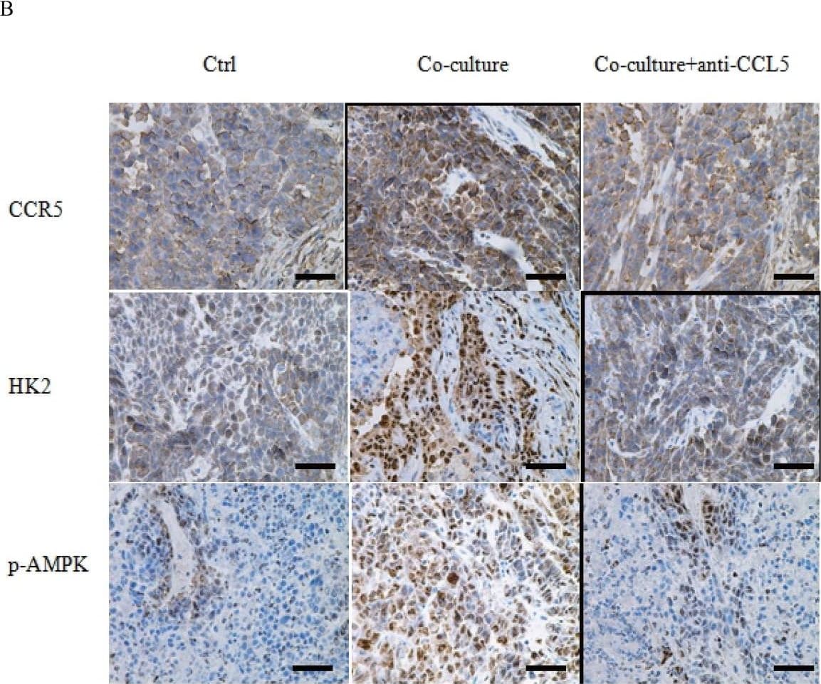
Detection of Human CCR5 by Immunohistochemistry Macrophages promoted breast cancer metastasis through CCL5(A) MDA-MB-231 cells were co-cultured with 15 mM lactate-activated THP-1 macrophages for 7 days, in the presence of 5μg/ml anti-CCL5 neutralizing antibody or not. MDA-MB-231 cells were then collected and injected into the tail vein of nude mice. After two weeks, animals were sacrificed and metastatic nodules on lung surfaces were counted. (B) CCR5, HK2 and p-AMPK were immunostained in MDA-MB-231 metastases. Scale bars represent 50 μm. *, P<0.05; **, P<0.01. Image collected and cropped by CiteAb from the following publication (https://www.oncotarget.com/lookup/doi/10.18632/oncotarget.22786), licensed under a CC-BY license. Not internally tested by R&D Systems.
 View Larger
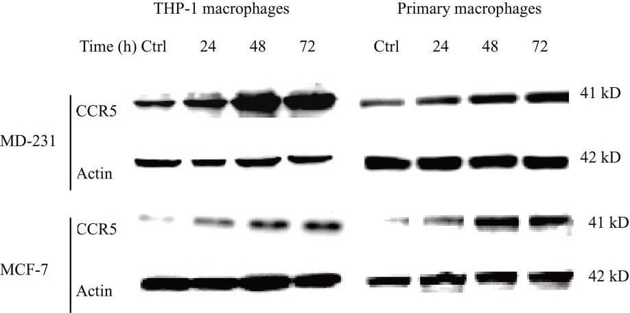
Detection of Human CCR5 by Western Blot Lactate-activated macrophages induced EMT in breast cancer cells through CCL5-CCR5 axis(A) 106 THP-1 macrophages were treated with 15 mM lactate for 72 h, and then cells were washed twice and fresh media were added. Macrophages were cultured for another 24 h and the conditional media (lactate CM) was collected. The effect of CM on breast cancer cell migration was measured by double chamber transwell assay. 5μg/ml anti-CCL5 neutralizing antibody significantly decreased lactate CM-induced cell migration. (B) 106 MCF-7 cells were co-cultured with 15 mM lactate-activated macrophages in the presence of 5μg/ml anti-CCL5 antibody or not, and protein levels of EMT markers were tested by western blot. (C) 106 breast cancer cells were co-cultured with 106 lactate-activated THP-1 macrophages (or 106 lactate-activated primary macrophages) for different time points, and the expression of CCR5 was monitored by western blot. (D) MDA-MB-231 and MCF-7 cells were transfected with shCCR5 plasmids, or pre-treated with 5μM Maraviroc for 2 h, then cell migration induced by lactate CM was detected by double chamber transwell assay. Lactate CM was described in (A). (E) MCF-7 cells (106) were transfected with pcDNA3.1-CCR5, and then cultured with 10ng/ml CCL5 for 24 h. The expression of E-cadherin, N-cadherin and vimentin was investigated by western blot. (F) 106 Human primary macrophages (No. 4 and No. 9) were treated with 15 mM lactate for 72 h and CM was collected as described in (A). The migration of MDA-MB-231 cells was measured in the presence of primary macrophage CM. 5μg/ml anti-CCL5 neutralizing antibody, shRNAs designed against CCR5, or 5μM Maraviroc, significantly reduced primary macrophage CM-induced cell migration. *, P<0.05; **, P<0.01. Image collected and cropped by CiteAb from the following publication (https://www.oncotarget.com/lookup/doi/10.18632/oncotarget.22786), licensed under a CC-BY license. Not internally tested by R&D Systems.
 View Larger
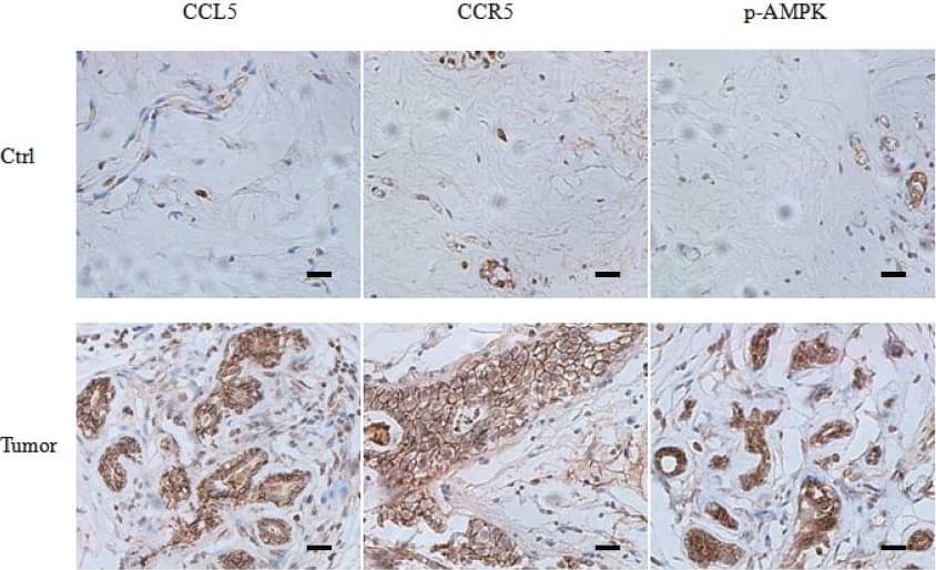
Detection of Human CCR5 by Immunohistochemistry CCL5-CCR5 axis induced aerobic glycolysis by regulation of AMPK signaling(A) Western blot for AMPK, c-Myc, HIF-1 alpha and Akt in breast cancer cells co-cultured with 15mM lactic acid-activated THP-1 macrophages (ratio 1:1) for 72 h. Results presented were representatives of at least three independent experiments. (B) The expression of AMPK downstream signaling target ACC in breast cancer cells co-cultured as in (A). (C) MDA-MB-231 and MCF-7 cells were transfected with 50 nM AMPK alpha 1 siRNA, or pretreated with 10μM compound C for 4 h, and then incubated with 15mM lactic acid-activated THP-1 macrophages (ratio 1:1) for 48 h. The glucose uptake, lactic acid production and ATP levels were detected. (D) The inhibition of AMPK abrogated macrophage-induced EMT in MCF-7 cells. Cells were treated as described in (C). After co-culture, the expression of EMT markers, E-cadherin and vimentin, was measured by western blot. (E) Recombinant human CCL5 induced the phosphorylation of AMPK in MDA-MB-231 and MCF-7/CCR5 cells. 106 cells were treated with 50ng/ml CCL5 for defferent time points as indicated, and phosphorylated AMPK and total AMPK were investigated by western blot. (F) Inhibition of CCR5 in MDA-MB-231 cells significantly attenuated macrophage-induced AMPK phosphorylation. MDA-MB-231 cells were transfected with shRNAs designed against CCR5, or pre-treated with 5μM Maraviroc for 2 h, then co-cultured with 15 mM lactate-activated macrophages as described in (A). After co-culture, the phosphorylation of AMPK was detected by western blot. (G) Expressions of CCL5, CCR5 and p-AMPK in samples obtained from breast cancer patients (n =28). Scale bars represent 50 μm. *, P<0.05; **, P<0.01. Image collected and cropped by CiteAb from the following publication (https://www.oncotarget.com/lookup/doi/10.18632/oncotarget.22786), licensed under a CC-BY license. Not internally tested by R&D Systems.
Human CCR5 Antibody Summary
Met1-Leu352
Accession # P51681
Applications
Please Note: Optimal dilutions should be determined by each laboratory for each application. General Protocols are available in the Technical Information section on our website.
Background: CCR5
CCR5 is a G protein-linked seven transmembrane domain chemokine receptor. CCR5 serves as a receptor for several chemokines including MIP-1 alpha, MIP-1 beta, MCP-2, and RANTES. It also functions as a coreceptor for Macrophage Tropic HIV-1 infection.
Preparation and Storage
- 12 months from date of receipt, -20 to -70 °C as supplied.
- 1 month, 2 to 8 °C under sterile conditions after reconstitution.
- 6 months, -20 to -70 °C under sterile conditions after reconstitution.
参考图片
CCR5 in Human Dorsal Root Ganglia. CCR5 was detected in immersion fixed paraffin-embedded sections of human dorsal root ganglia using 25 µg/mL Human CCR5 Monoclonal Antibody (Catalog # MAB181) overnight at 4 °C. Tissue was stained with the Anti-Mouse HRP-AEC Cell & Tissue Staining Kit (red; Catalog # CTS003) and counterstained with hematoxylin (blue). View our protocol for Chromogenic IHC Staining of Paraffin-embedded Tissue Sections.

 危险品化学品经营许可证(不带存储) 许可证编号:沪(杨)应急管危经许[2022]202944(QY)
  营业执照(三证合一)