


 用小程序,查商品更便捷


Functional Assay
1:500FC
1:500-1000
This antibody recognizes the same epitope as clone 15E8.
CD28 (Cluster of Differentiation 28) is one of the proteins expressed on T cells that provide co-stimulatory signals required for T cell activation and survival. T cell stimulation through CD28 in addition to the T-cell receptor (TCR) can provide a potent signal for the production of various interleukins (IL-6 in particular).
CD28 is the receptor for CD80 (B7.1) and CD86 (B7.2) proteins. When activated by Toll-like receptor ligands, the CD80 expression is upregulated in antigen-presenting cells (APCs). The CD86 expression on antigen-presenting cells is constitutive (expression is independent of environmental factors). It is generally reported, that CD28 is expressed on 50% of CD8+ T cells and more than 80% CD4+ T cells in human, but during the course of activation some T cells lose this molecule. In general, CD28 is a primary costimulatory molecule for T cell activation.
12 months from date of receipt / reconstitution, 2 to 8 °C as supplied
参考图片
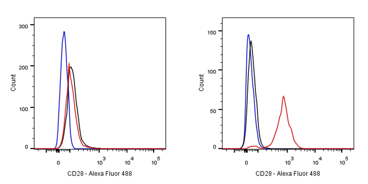
Flow cytometric analysis of THP-1 (Human monocytic leukemia monocyte, left) / Jurkat (Human T cell leukemia T lymphocyte, right) cells labelling CD28 antibody at 1/500 dilution (0.1 μg)/ (red) compared with a Rabbit monoclonal IgG (Black) isotype control and an unlabelled control (cells without incubation with primary antibody and secondary antibody) (Blue). Goat Anti-Mouse IgG Alexa Fluor® 488 was used as the secondary antibody. Negative control: THP-1
T cell proliferation Assay with CD3(S0B0009) &CD28 (S0B0010)
T cell proliferation Assay with CD3(S0B0009) &CD28 (S0B0010)
Procedure
1. Isolate human PBMCs according to the manufacturer’s instructions.
2. Suspend PBMCs in medium at 106 cells/mL.
3. Do one of the following depending on the format you are using:
· For the soluble format, incubate 106 cells with CD3 Mouse mAb (OKT3) at 1 µg/mL final concentration in culture for 3 days (5% CO2, 37°C).
· For the immobilized format, coat the CD3 Mouse mAb (OKT3) onto a plate at 5 to 10 μg/mL at 4°C, overnight. Add cells to the CD3 Mouse mAb (OKT3)-coated plate.
4. Wash the cells twice and resuspend them in medium at 106 cells/mL.
5. Distribute the cells in a round-bottom plate at 105 cells per well.
6. [Optional for co-stimulation] Add CD28 Mouse mAb (15E8) and optionally Protein G at 5 µg/mL each final concentration, and incubate the plate for an additional 3 days (5% CO2 at 37°C).

 危险品化学品经营许可证(不带存储) 许可证编号:沪(杨)应急管危经许[2022]202944(QY)
  营业执照(三证合一)