


 用小程序,查商品更便捷


Product Usage Information
| Application | Dilution |
|---|---|
| Immunohistochemistry (Paraffin) | 1:200 - 1:800 |
| Immunofluorescence (Immunocytochemistry) | 1:400 - 1:800 |
| Flow Cytometry (Fixed/Permeabilized) | 1:200 - 1:800 |
| Flow Cytometry (Live) | 1:200 - 1:800 |
Specificity/Sensitivity
物种反应性:
人, 猴
参考图片
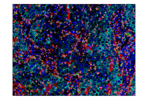
Confocal immunofluorescent analysis of THP-1 (left) and Jurkat (right) cells using CD68 (D4B9C) XP® Rabbit mAb (green). Blue pseudocolor = DRAQ5® #4084 (fluorescent DNA dye). Projected images from a z-stack series are shown.
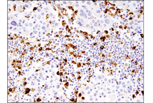
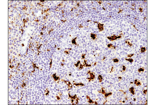
Immunohistochemical analysis of paraffin-embedded human tonsil using CD68 (D4B9C) XP® Rabbit mAb.
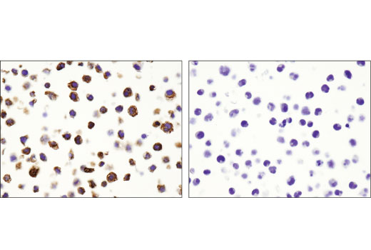
Immunohistochemical analysis of paraffin-embedded THP-1 (left) and Jurkat (right) cell pellets using CD68 (D4B9C) XP® Rabbit mAb.
Flow cytometric analysis of human peripheral blood mononuclear cells co-stained with anti-human CD14 using CD68 (D4B9C) XP® Rabbit mAb (right) compared to a concentration-matched Rabbit (DA1E) mAb IgG XP® Isotype Control #3900 (left). Anti-rabbit IgG (H+L), F(ab')2 Fragment (Alexa Fluor® 488 Conjugate) #4412 was used as a secondary antibody.
Flow cytometric analysis of Jurkat cells (blue) and THP-1 cells (green) using CD68 (D4B9C) XP® Rabbit mAb. Anti-rabbit IgG (H+L), F(ab')2 Fragment (Alexa Fluor® 488 Conjugate) #4412 was used as a secondary antibody.
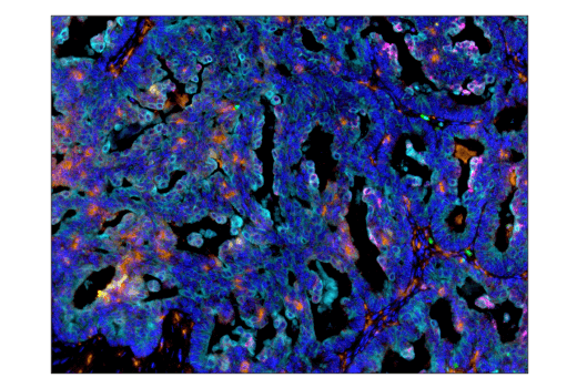
Immunohistochemical analysis of paraffin-embedded human serous papillary carcinoma of the ovary using CD68 (D4B9C) XP® Rabbit mAb.
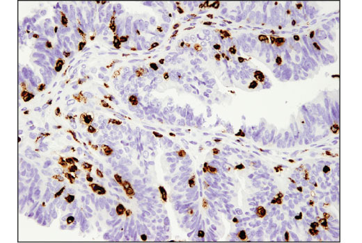
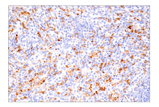
Immunohistochemical analysis of paraffin-embedded human lung carcinoma using CD68 (D4B9C) XP® Rabbit mAb.

 危险品化学品经营许可证(不带存储) 许可证编号:沪(杨)应急管危经许[2022]202944(QY)
  营业执照(三证合一)