


1/3
BD Horizon™ BV605 Mouse Anti-Human CD8


品牌: BD Pharmingen
 下载产品说明书 下载SDS
 用小程序,查商品更便捷


 收藏
收藏
 对比
对比 咨询
咨询反应种属:
Human (QC Testing), Rhesus,Cynomolgus,Baboon (Tested in Development)
Human (QC Testing), Rhesus,Cynomolgus,Baboon (Tested in Development)
来源宿主:
Mouse BALB/c IgG1, κ
Mouse BALB/c IgG1, κ
产品介绍
产品信息
耦联标记
BV605

抗原名称
CD8
宿主
Mouse BALB/c IgG1, κ
免疫原
Human Peripheral Blood T Cells
简单描述
The SK1 monoclonal antibody specifically binds to CD8 alpha (CD8α). CD8α is a type I transmembrane glycoprotein and a member of the immunoglobulin superfamily. CD8α is expressed by the majority of thymocytes, by subpopulations of αβ T cells and γδ T cells and by some NK cells. Cell surface CD8α is expressed either as a disulfide-linked homodimer (CD8αα) or as a heterodimer (CD8αβ) when disulfide-bonded to a CD8 beta chain (CD8β). CD8-positive αβ T cells coexpress both CD8αα homodimers and CD8αβ heterodimers whereas som
e γδ T cells and NK cells express CD8αα homodimers. CD8 plays important roles in T cell activation and selection. The extracellular IgSF domain of CD8α binds to a non-polymorphic determinant on HLA class I molecules (α3 domain) and enables CD8 to function as a co-receptor with MHC class I-restricted TCR during T cell recognition of antigen. The cytoplasmic domain of CD8α associates with Lck, a Src family protein tyrosine kinase that is involved in intracellular signaling.
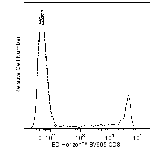
This antibody is conjugated to BD Horizon BV605 which is part of the BD Horizon Brilliant™ Violet family of dyes. With an Ex Max of 407-nm and Em Max of 602-nm, BD Horizon BV605 can be excited by a violet laser and detected with a standard 610/20-nm filter set. BD Horizon BV605 is a tandem fluorochrome of BD Horizon BV421 and an acceptor dye with an Em max at 605-nm. Due to the excitation of the acceptor dye by the green (532 nm) and yellow-green (561 nm) lasers, there will be significant spillover into the PE and BD Horizon PE-CF594 detectors off the green or yellow-green lasers. BD Horizon BV605 conjugates are very bright, often exhibiting brightness equivalent to PE conjugates and can be used as a third color off of the violet laser.
For optimal and reproducible results, BD Horizon Brilliant Stain Buffer should be used anytime two or more BD Horizon Brilliant dyes are used in the same experiment. Fluorescent dye interactions may cause staining artifacts which may affect data interpretation. The BD Horizon Brilliant Stain Buffer was designed to minimize these interactions. More information can be found in the Technical Data Sheet of the BD Horizon Brilliant Stain Buffer (Cat. No. 563794).
商品描述
The SK1 monoclonal antibody specifically binds to CD8 alpha (CD8α). CD8α is a type I transmembrane glycoprotein and a member of the immunoglobulin superfamily. CD8α is expressed by the majority of thymocytes, by subpopulations of αβ T cells and γδ T cells and by some NK cells. Cell surface CD8α is expressed either as a disulfide-linked homodimer (CD8αα) or as a heterodimer (CD8αβ) when disulfide-bonded to a CD8 beta chain (CD8β). CD8-positive αβ T cells coexpress both CD8αα homodimers and CD8αβ heterodimers whereas som
e γδ T cells and NK cells express CD8αα homodimers. CD8 plays important roles in T cell activation and selection. The extracellular IgSF domain of CD8α binds to a non-polymorphic determinant on HLA class I molecules (α3 domain) and enables CD8 to function as a co-receptor with MHC class I-restricted TCR during T cell recognition of antigen. The cytoplasmic domain of CD8α associates with Lck, a Src family protein tyrosine kinase that is involved in intracellular signaling.
This antibody is conjugated to BD Horizon BV605 which is part of the BD Horizon Brilliant™ Violet family of dyes. With an Ex Max of 407-nm and Em Max of 602-nm, BD Horizon BV605 can be excited by a violet laser and detected with a standard 610/20-nm filter set. BD Horizon BV605 is a tandem fluorochrome of BD Horizon BV421 and an acceptor dye with an Em max at 605-nm. Due to the excitation of the acceptor dye by the green (532 nm) and yellow-green (561 nm) lasers, there will be significant spillover into the PE and BD Horizon PE-CF594 detectors off the green or yellow-green lasers. BD Horizon BV605 conjugates are very bright, often exhibiting brightness equivalent to PE conjugates and can be used as a third color off of the violet laser.
For optimal and reproducible results, BD Horizon Brilliant Stain Buffer should be used anytime two or more BD Horizon Brilliant dyes are used in the same experiment. Fluorescent dye interactions may cause staining artifacts which may affect data interpretation. The BD Horizon Brilliant Stain Buffer was designed to minimize these interactions. More information can be found in the Technical Data Sheet of the BD Horizon Brilliant Stain Buffer (Cat. No. 563794).
同种型
Mouse BALB/c IgG1, κ
克隆号
克隆 SK1 (also known as Leu-2a; Leu2a) (RUO)
产品详情
BV605
The BD Horizon Brilliant Violet™ 605 (BV605) dye is part of the BD Horizon Brilliant Violet™ family of dyes. This tandem fluorochrome is comprised of a BV421 donor with an excitation maximum (Ex Max) of 407-nm and an acceptor dye with an emission maximum (Em Max) at 605-nm. BV605, driven by BD innovation, is designed to be excited by the violet laser (405-nm) and detected using an optical filter centered near 610-nm (e.g., a 610/20-nm bandpass filter). The acceptor dye can be excited by the yellow-green (561-nm) laser resulting in cross-laser excitation and fluorescence spillover. Please ensure that your instrument’s configurations (lasers and optical filters) are appropriate for this dye.

BV605
Violet 405 nm
407 nm
605 nm
应用
实验应用
Flow cytometry (Routinely Tested)
推荐用量
5 µl
反应种属
Human (QC Testing), Rhesus,Cynomolgus,Baboon (Tested in Development)
目标/特异性
CD8
背景
别名
CD8α; CD8A; CD8 alpha; Leu2a; MAL; T8; p32
制备和贮存
存储溶液
Aqueous buffered solution containing ≤0.09% sodium azide.
保存方式
Aqueous buffered solution containing BSA and ≤0.09% sodium azide.
文献
文献
研发参考(9)
1. Bernard A, Boumsell L, Hill C. Joint report of the first international workshop on human leucocyte differentiation antigens by the investigators of the participating laboratories: T2 protocol. In: Bernard A. A. Bernard .. et al., ed. Leucocyte typing : human leucocyte differentiation antigens detected by monoclonal antibodies : specification, classification, nomenclature = Typage leucocytaire : antigènes de différenciation leucocytaire humains révélés par les anticorps monoclonaux : "Rapports des études communes". Berlin New York: Springer-Verlag; 1984:25-60.
2. Dongworth DW, Gotch FM, Carter NP, Hildreth PDK, McMichael AJ. Inhibition of virus-specific, HLA-restricted, T cell-mediated lysis by monoclonal anti-T cell antibodies. In: Bernard A. A. Bernard .. et al., ed. Leucocyte typing : human leucocyte differentiation antigens detected by monoclonal antibodies : specification, classification, nomenclature = Typage leucocytaire : antigènes de différenciation leucocytaire humains révélés par les anticorps monoclonaux : "Rapports des études communes". Berlin New York: Springer-Verlag; 1984:320-328.
3. Engleman EG, Benike CJ, Glickman E, Evans RL. Antibodies to membrane structures that distinguish suppressor/cytotoxic and helper T lymphocyte subpopulations block the mixed leukocyte reaction in man. J Exp Med. 1981; 154(1):193-198. (Clone-specific: Cell separation, Flow cytometry, Functional assay, Inhibition).
4. Engleman EG, Benike CJ, Grumet FC, Evans RL. Activation of human T lymphocyte subsets: helper and suppressor/cytotoxic T cells recognize and respond to distinct histocompatibility antigens. J Immunol. 1981; 127(5):2124-2129. (Clone-specific: Cell separation, Flow cytometry, Fluorescence activated cell sorting).
5. Evans RL, Wall DW, Platsoucas CD, et al. Thymus-dependent membrane antigens in man: inhibition of cell-mediated lympholysis by monoclonal antibodies to TH2 antigen. Proc Natl Acad Sci U S A. 1981; 78(1):544-548. (Immunogen: Flow cytometry, Functional assay, Inhibition).
6. Jonker M, Meurs G. Monoclonal antibodies specific for B cells, cytotoxic/suppressor T cells, and a subset of cytotoxic/suppressor T cells in the Rhesus monkey. In: Bernard A. A. Bernard .. et al., ed. Leucocyte typing : human leucocyte differentiation antigens detected by monoclonal antibodies : specification, classification, nomenclature = Typage leucocytaire : antigènes de différenciation leucocytaire humains révélés par les anticorps monoclonaux : "Rapports des études communes". Berlin New York: Springer-Verlag; 1984:328-336.
7. Ledbetter JA, Evans RL, Lipinski M, Cunningham-Rundles C, Good RA, Herzenberg LA. Evolutionary conservation of surface molecules that distinguish T lymphocyte helper/inducer and cytotoxic/suppressor subpopulations in mouse and man. J Exp Med. 1981; 153(2):310-323. (Clone-specific: Flow cytometry, Immunoprecipitation).
8. McMichael AJ. A.J. McMichael .. et al., ed. Leucocyte typing III : white cell differentiation antigens. Oxford New York: Oxford University Press; 1987:1-1050.
9. Warner NL, Lanier LL, Jackson A, Babcock G, Evans R. Multiparameter approaches to FACS analysis of human leucocyte cell surface antigens. In: Bernard A. A. Bernard .. et al., ed. Leucocyte typing : human leucocyte differentiation antigens detected by monoclonal antibodies. Berlin New York: Springer-Verlag; 1984:621-630.
声明 :本官网所有报价均为常温或者蓝冰运输价格,如有产品需要干冰运输,需另外加收干冰运输费。

 危险品化学品经营许可证(不带存储) 许可证编号:沪(杨)应急管危经许[2022]202944(QY)
  营业执照(三证合一)