


 用小程序,查商品更便捷


WB
1:1000IP
1:50IHC-P
1:500ICC
1:500
The epidermal growth factor receptor (EGFR; ErbB-1; HER1 in humans) is a transmembrane protein that is a receptor for members of the epidermal growth factor family (EGF family) of extracellular protein ligands. The EGFR is essential for ductal development of the mammary glands, and agonists of the EGFR such as amphiregulin, TGF-α, and heregulin induce both ductal and lobuloalveolar development even in the absence of estrogen and progesterone. EGFR mutations, del E746-A750 in exon 19 and L858R in exon 21 in tumor cells of NMLC represent biomarkers of response to tyrosine kinase inhibitors (TKI) therapy. Patients with tumors positive for EGFR mutations show better response and greater survival. These mutations occupy 90% of mutations in lung cancer.
12 months from date of receipt / reconstitution, -20 °C as supplied
参考图片
WB result of EGFR (delE746-A750) Rabbit mAb
Primary antibody: EGFR (delE746-A750) Rabbit mAb at 1/1000 dilution
Lane 1: A431 whole cell lysate 20 µg
Lane 2: A549 whole cell lysate 20 µg
Lane 3: H1975 whole cell lysate 20 µg
Lane 4: HCC827 whole cell lysate 20 µg
Negative control: A431 whole cell lysate; A549 whole cell lysate; H1975 whole cell lysate
Secondary antibody: Goat Anti-Rabbit IgG, (H+L), HRP conjugated at 1/10000 dilution
Predicted MW: 134 kDa
Observed MW: 175 kDa
EGFR (delE746-A750) Rabbit mAb at 1/50 dilution (1 µg) immunoprecipitating EGFR (delE746-A750) in 0.4 mg HCC827 whole cell lysate.
Western blot was performed on the immunoprecipitate using EGFR (delE746-A750) Rabbit mAb at 1/1000 dilution.
Secondary antibody (HRP) for IP was used at 1/400 dilution.
Lane 1: HCC827 whole cell lysate 20 µg (Input)
Lane 2: EGFR (delE746-A750) Rabbit mAb IP in HCC827 whole cell lysate
Lane 3: Rabbit monoclonal IgG IP in HCC827 whole cell lysate
Predicted MW: 134 kDa
Observed MW: 175 kDa
IHC shows positive staining in paraffin-embedded HCC827 cells and negative staining in paraffin-embedded A549, H1975 and A431 cells. Anti-EGFR (delE746-A750) antibody was used at 1/500 dilution, followed by a HRP Polymer for Mouse & Rabbit IgG (ready to use). Counterstained with hematoxylin. Heat mediated antigen retrieval with Tris/EDTA buffer pH9.0 was performed before commencing with IHC staining protocol.
IHC shows positive staining in paraffin-embedded human lung adenocarcinoma. Anti-EGFR (delE746-A750) antibody was used at 1/500 dilution, followed by a HRP Polymer for Mouse & Rabbit IgG (ready to use). Counterstained with hematoxylin. Heat mediated antigen retrieval with Tris/EDTA buffer pH9.0 was performed before commencing with IHC staining protocol.
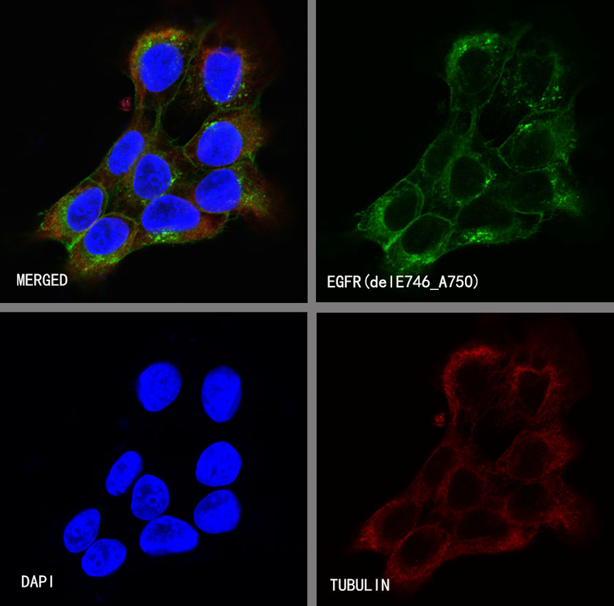
ICC shows positive staining in HCC827 cells. Anti-EGFR(delE746_A750) antibody was used at 1/500 dilution (Green) and incubated overnight at 4°C. Goat polyclonal Antibody to Rabbit IgG - H&L (Alexa Fluor® 488) was used as secondary antibody at 1/1000 dilution. The cells were fixed with 100% ice-cold methanol and permeabilized with 0.1% PBS-Triton X-100. Nuclei were counterstained with DAPI (Blue).Counterstain with tubulin (red).
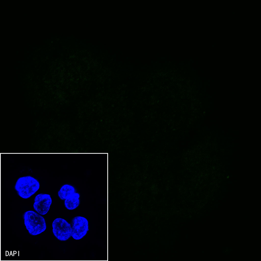
Negative control:ICC shows negative staining in A431 cells. Anti-EGFR(delE746_A750) antibody was used at 1/500 dilution and incubated overnight at 4°C. Goat polyclonal Antibody to Rabbit IgG - H&L (Alexa Fluor® 488) was used as secondary antibody at 1/1000 dilution. The cells were fixed with 100% ice-cold methanol and permeabilized with 0.1% PBS-Triton X-100. Nuclei were counterstained with DAPI (Blue).

 危险品化学品经营许可证(不带存储) 许可证编号:沪(杨)应急管危经许[2022]202944(QY)
  营业执照(三证合一)