


 下载产品说明书 下载SDS
 用小程序,查商品更便捷


 收藏
收藏
 对比
对比 咨询
咨询

参考图片
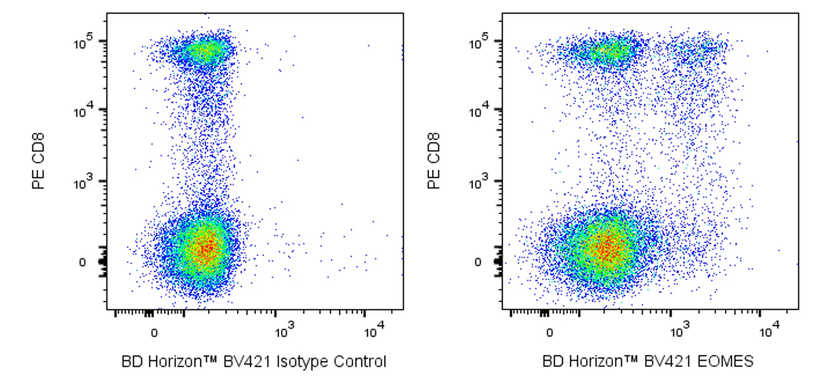
Flow cytometric analysis of EOMES expression in human peripheral blood lymphocytes. Peripheral blood mononuclear cells were stained intracellularly with PE Mouse Anti-Human CD8 antibody (Cat. No. 555367/557086/561949) and either BD Horizon™ BV421 Mouse IgG1, κ Isotype Control (Cat. No. 562438; Left Plot) or BD Horizon BV421 Mouse Anti-EOMES antibody (Cat. No. 567166; Right Plot) at 0.5 µg/test using BD Pharmingen™ Transcription Factor Buffer Set (Cat. No. 562574/562725). The bivariate pseudocolor density plot showing the correlated expression of EOMES (or Ig Isotype control staining) versus CD8 was derived from events with the forward and side light-scatter characteristics of intact lymphocytes. Flow cytometry and data analysis was performed using a BD LSRFortessa™ Flow Cytometer System and FlowJo™ software. Data shown on this Technical Data Sheet are not lot specific.
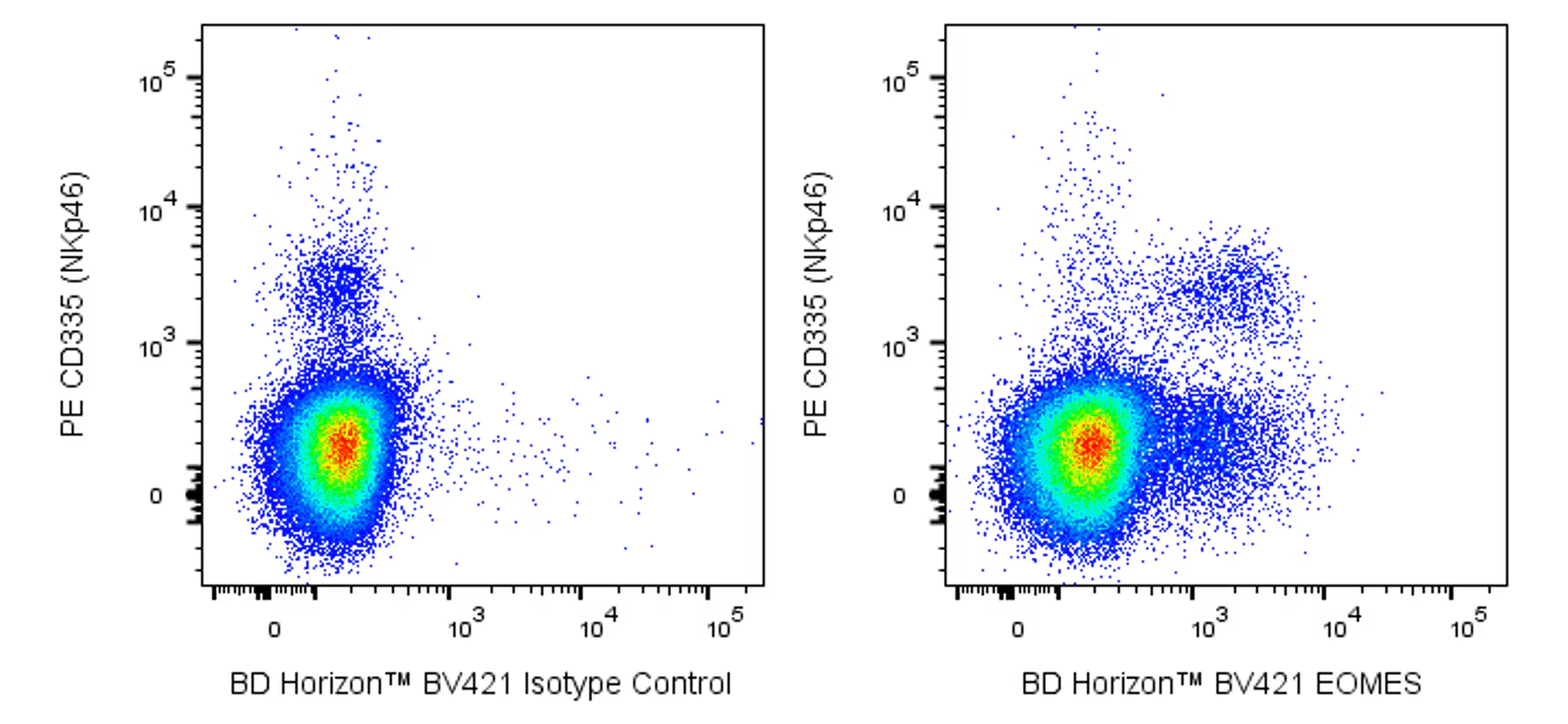
Flow cytometric analysis of EOMES expression in mouse splenic leucocytes. Spleen cells from C57BL/6 mice were stained intracellularly with PE Rat Anti-Mouse CD335 (NKp46) antibody (Cat.No. 560757) and either BD Horizon™ BV421 Mouse IgG1, κ Isotype Control (Left Plot) or BD Horizon BV421 Mouse Anti-EOMES antibody (Right Plot) at 0.5 µg/test using BD Pharmingen™ Transcription Factor Buffer Set. The bivariate pseudocolor density plot showing the correlated expression of EOMES (or Ig isotype control staining) versus CD355 (NKp46) were derived from events with the forward and side light-scatter characteristics of intact leucocytes. Flow cytometry and data analysis was performed using a BD LSRFortessa™ Flow Cytometer System and FlowJo™ software. Data shown on this Technical Data Sheet are not lot specific.

 危险品化学品经营许可证(不带存储) 许可证编号:沪(杨)应急管危经许[2022]202944(QY)
  营业执照(三证合一)