ER Stress-induced Autophagy Antibody Sampler Kit
1/26

ER Stress-induced Autophagy Antibody Sampler Kit

分享
品牌: CST
pdf 下载产品说明书
用小程序,查商品更便捷
收藏
对比对比
咨询咨询
    产品介绍
    产品信息
    抗原名称
    ER Stress-induced Autophagy
    来源纯化
    Monoclonal antibody is produced by immunizing animals with a synthetic peptide corresponding to residues surrounding Gly584 of human BiP, residues surrounding Ser36 of human Atg12 protein, residues surrounding Thr72 of human Beclin-1, residues surrounding Ser51 of human eIF2α, residues surrounding Thr183/Tyr185 of human SAPK/JNK, residues of a purified recombinant human eIF2α, and residues corresponding to the amino terminus of human JNK1.
    简单描述
    Antibody Sampler Kit for studying SAPK/JNK1/SAPK/JNK1 (Thr183/Tyr185) phosphate/EIF2S1/eIF2-alpha/EIF2S1/eIF2-alpha (Ser52) phosphate/BiP/Beclin 1/ATG12 in the research area.
    研究领域
    癌症,细胞生物学,纤维化,代谢,神经科学,RNA 调节与翻译调控
    应用
    目标/特异性

    Specificity/Sensitivity

    Each antibody in the ER Stress-induced Antibody Sampler Kit detects endogenous levels of its target protein. Phospho-eIF2α (Ser51) (D9G8) XP® Rabbit mAb detects endogenous eIF2α only when phosphorylated at Ser51. The antibody does not recognize elF2α phosphorylated at other sites. Phospho-SAPK/JNK (Thr183/Tyr185) (81E11) Rabbit mAb detects endogenous levels of p46 and p54 SAPK/JNK only when phosphorylated at Thr183 and Tyr185. This antibody may cross-react with phosphorylated p44/42 or p38 MAP kinases. JNK1 (2C6) Mouse mAb detects endogenous levels of total JNK1 protein. This antibody may cross react with recombinant levels JNK2 protein. The antibody does not cross react with JNK3 protein.

    背景
    背景
    The endoplasmic reticulum (ER) is an organelle with essential biosynthetic and signaling functions in eukaryotic cells (1). Post synthesis of secretory and transmembrane proteins on polysomes, proteins are translocated into the ER where they are often modified by disulfide bond formation, amino-linked glycosylation, and folding. Different physiological and pathological conditions can disturb proper protein folding in the ER causing ER stress (1). ER stress activates an intracellular signaling transduction pathway called unfolded protein response (UPR) and autophagy to avoid cell death (2). The main role of UPR is to improve the protein load on the ER by shutting down protein translation and gene transcription to enhance ER's folding capacity (2). On the other hand, autophagy is a catabolic process for the autophagosomic-lysosomal degradation of bulk cytoplasmc contents (3,4). One of the chaperones aiding in proper protein folding is Binding immunoglobulin Protein (BiP) (5,6). BiP works by binding to misfolded proteins to prevent them from forming aggregates and assists in proper refolding (7). The molecular machinery of autophagy was largely discovered in yeast and referred to as autophagy-related (Atg) genes. Formation of the autophagosome involves a ubiquitin-like conjugation system in which Atg12 is covalently bound to Atg5 and targeted to autophagosome vesicles (8-10). One of the proteins critical to autophagy process is Beclin-1, the mammalian orthologue of the yeast autophagy protein Apg6/Vps30 (11). Beclin-1 can complement defects in yeast autophagy caused by loss of Apg6 and can also stimulate autophagy when overexpressed in mammalian cells (12). Mammalian Beclin-1 was originally isolated in a yeast two-hybrid screen for Bcl-2 interacting proteins and has been shown to interact with Bcl-2 and Bcl-xL, but not with Bax or Bak (13). Phosphorylation of the eukaryotic initiation factor 2 (eIF2) α subunit is a well-documented mechanism to downregulate protein synthesis under a variety of stress conditions. eIF2 binds GTP and Met-tRNAi and transfers Met-tRNA to the 40S subunit to form the 43S preinitiation complex (14,15). Kinases that are activated by viral infection (PKR) can phosphorylate the α subunit of eIF2 (16,17). Induction of PKR by IFN-γ and TNF-α induces potent phosphorylation of eIF2α at Ser51 (18,19). There are three SAPK/JNK genes each of which undergoes alternative splicing, resulting in numerous isoforms (20). The IRE1, a transmembrane serine/threonine kinase (21,22), through its kinase activity activates SAPK/JNK in the early stage of ER stress in order to induce autophagosome formation (23). 1.Verfaillie, T. et al. (2010) Int J Cell Biol 2010, 930509. 2.Ogata, M. et al. (2006) Mol Cell Biol 26, 9220-31. 3.Reggiori, F. and Klionsky, D.J. (2002) Eukaryot Cell 1, 11-21. 4.Codogno, P. and Meijer, A.J. (2005) Cell Death Differ 12 Suppl 2, 1509-18. 5.Wabl, M. and Steinberg, C. (1982) Proc Natl Acad Sci U S A 79, 6976-8. 6.Haas, I.G. and Wabl, M. (2002) Nature 306, 387-9. 7.Kohno, K. et al. (1993) Mol Cell Biol 13, 877-90. 8.Mizushima, N. et al. (1998) J Biol Chem 273, 33889-92. 9.Mizushima, N. et al. (1998) Nature 395, 395-8. 10.Suzuki, K. et al. (2001) EMBO J 20, 5971-81. 11.Kametaka, S. et al. (1998) J Biol Chem 273, 22284-91. 12.Liang, X.H. et al. (1999) Nature 402, 672-6. 13.Liang, X.H. et al. (1998) J Virol 72, 8586-96. 14.Kimball, S.R. (1999) Int J Biochem Cell Biol 31, 25-9. 15.de Haro, C. et al. (1996) FASEB J 10, 1378-87. 16.Kaufman, R.J. (1999) Genes Dev 13, 1211-33. 17.Sheikh, M.S. and Fornace, A.J. (1999) Oncogene 18, 6121-8. 18.Cheshire, J.L. et al. (1999) J Biol Chem 274, 4801-6. 19.Zamanian-Daryoush, M. et al. (2000) Mol Cell Biol 20, 1278-90. 20.Kyriakis, J.M. and Avruch, J. (2001) Physiol Rev 81, 807-69. 21.Nikawa, J. and Yamashita, S. (1992) Mol Microbiol 6, 1441-6. 22.Cox, J.S. et al. (1993) Cell 73, 1197-206. 23.Urano, F. et al. (2000) Science 287, 664-6.
    研究领域
    癌症,细胞生物学,纤维化,代谢,神经科学,RNA 调节与翻译调控
    数据库链接
    Entrez-Gene ID
    5599,1965,3309,8678,9140
    UniProt ID
    P45983,P05198,P11021,Q14457,O94817

    参考图片

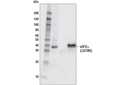
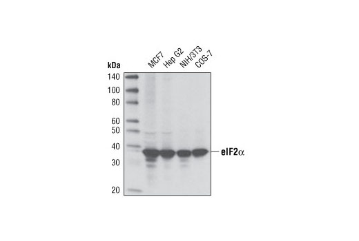
    Western blot analysis of extracts from various cell lines using eIF2α (D7D3) XP® Rabbit mAb.

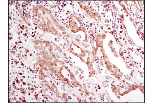
    Immunohistochemical analysis of paraffin-embedded human lung carcinoma using eIF2α (D7D3) XP® Rabbit mAb.

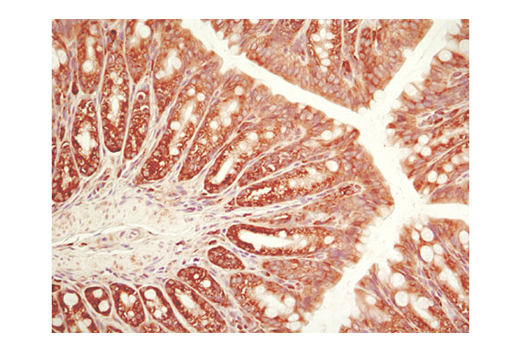
    Immunohistochemical analysis of paraffin-embedded mouse colon using eIF2α (D7D3) XP® Rabbit mAb.

    After the primary antibody is bound to the target protein, a complex with HRP-linked secondary antibody is formed. The LumiGLO® is added and emits light during enzyme catalyzed decomposition.

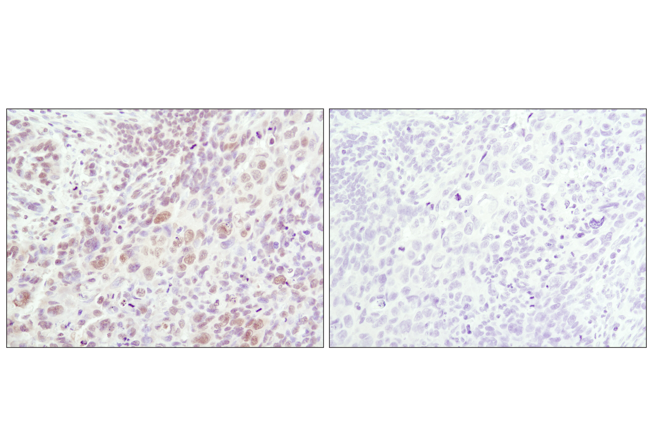
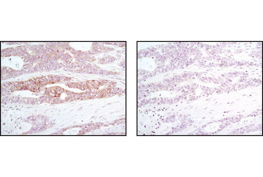
    Immunohistochemical analysis of paraffin-embedded human breast carcinoma using BiP (C50B12) Rabbit mAb in the presence of control peptide (left) or BiP Blocking Peptide #1084 (right).

    Immunohistochemical analysis of paraffin-embedded human lung carcinoma using Phospho-SAPK/JNK (Thr183/Tyr185) (81E11) Rabbit mAb in the presence of control peptide (left) or Phospho-SAPK/JNK (Thr183/Tyr185) Blocking Peptide #1215 (right).

    Immunohistochemical analysis of paraffin-embedded 293T cells untreated (left) or UV-treated (right) using Phospho-SAPK/JNK (Thr183/Tyr185) (81E11) Rabbit mAb.

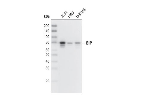
    Western blot analysis of extracts from various cell lines using BiP (C50B12) Rabbit mAb.

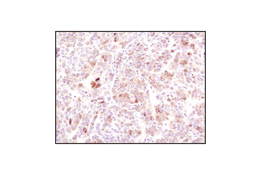
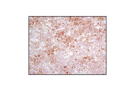
    Immunohistochemical analysis of paraffin-embedded human glioblastoma using BiP (C50B12) Rabbit mAb.

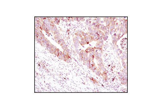
    Immunohistochemical analysis of paraffin-embedded human colon carcinoma using BiP (C50B12) Rabbit mAb.

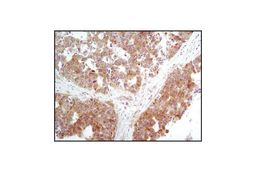
    Immunohistochemical analysis of paraffin-embedded human hepatocellular carcinoma using BiP (C50B12) Rabbit mAb.

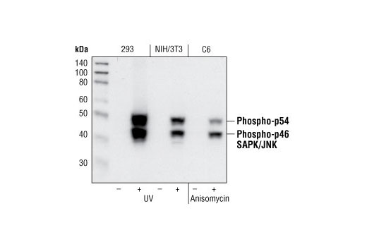
    Western blot analysis of extracts from 293 cells, untreated or UV-treated, NIH/3T3 cells, untreated or UV-treated and C6 cells, untreated or anisomycin-treated, using Phospho-SAPK/JNK (Thr183/Tyr185) (81E11) Rabbit mAb.

    Immunohistochemical analysis of frozen SKOV-3 xenograft using BiP (C50B12) Rabbit mAb.

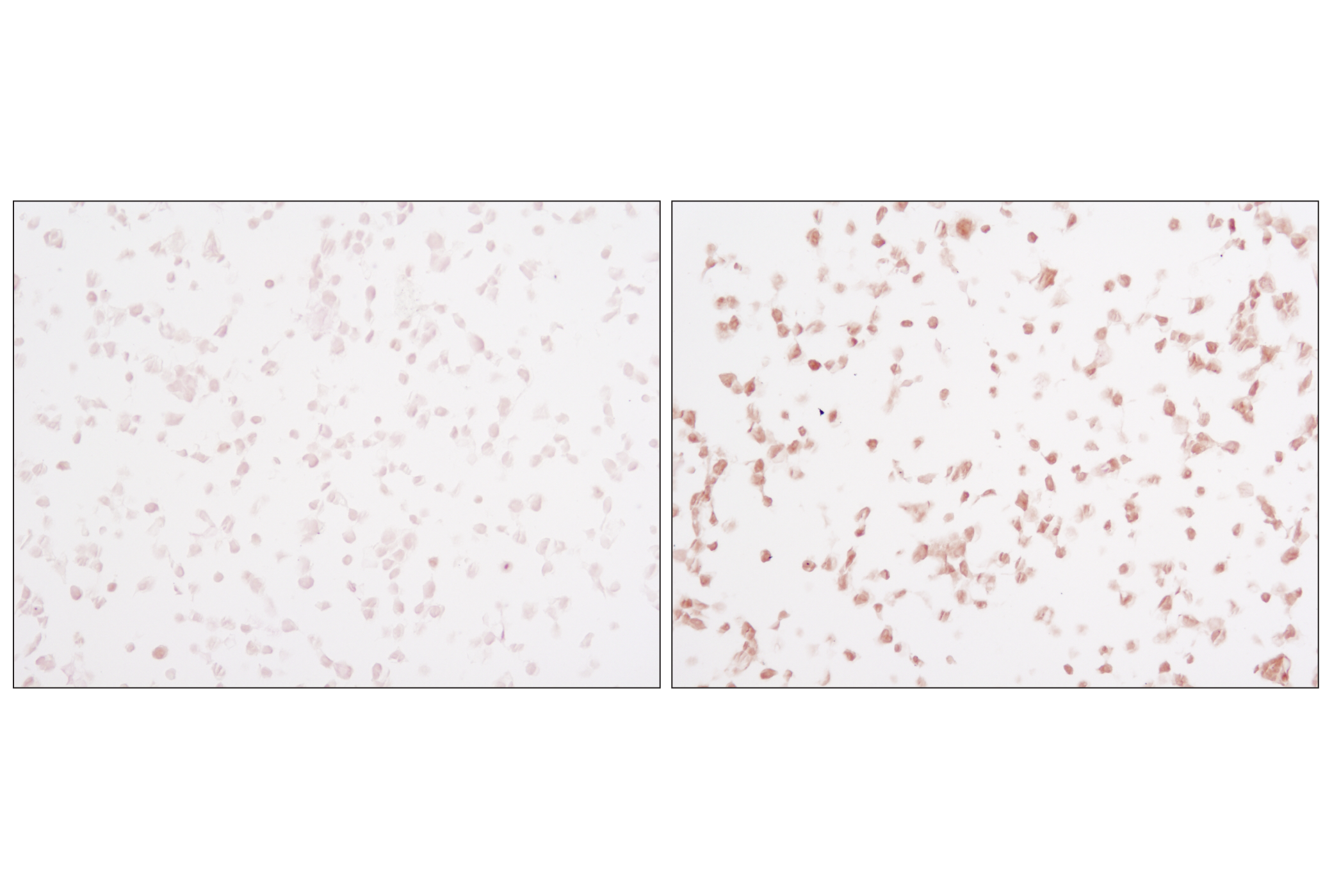
    Immunohistochemical analysis of paraffin-embedded human colon carcinoma, untreated (left) or λ phosphatase-treated (right), using Phopsho-eIF2α (Ser51) (D9G8) XP® Rabbit mAb.

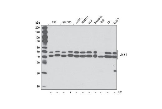
    Western blot analysis of extracts from indicated cell lines, untreated or UV-treated (40 J/m2, 30 min recovery), using JNK1 (2C6) Mouse mAb.

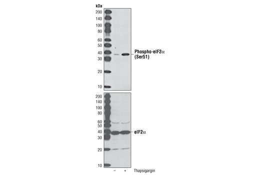
    Western blot analysis of extracts from C2C12 cells, untreated or thapsigargin-treated, using Phospho-eIF2α (Ser51) (D9G8) XP® Rabbit mAb (upper) or eIF2α Antibody #9722 (lower).

    Immunohistochemical analysis of paraffin-embedded human lung carcinoma using Phospho-eIF2α (Ser51) (D9G8) XP® Rabbit mAb.

    Immunohistochemical analysis of paraffin-embedded human lymphoma using Phospho-eIF2α (Ser51) (D9G8) XP® Rabbit mAb.

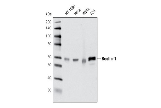
    Western blot analysis of extracts from various cell lines using Beclin-1 (D40C5) Rabbit mAb.

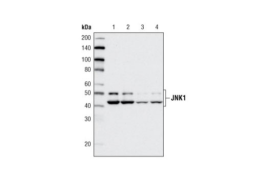
    Western blot analysis of HeLa cell extracts, untransfected (lane 1), mock-transfected (lane 2) or transfected with SignalSilence® SAPK/JNK siRNA I #6232 (lane 3) or SignalSilence® SAPK/JNK siRNA II #6233 (lane 4) for 72 hours, using JNK1 (2C6) Mouse mAb.

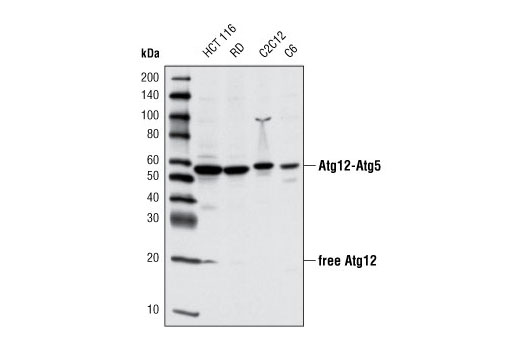
    Western blot analysis of extracts from various cell lines using Atg12 (D88H11) Rabbit mAb.

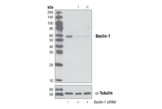
    Western blot analysis of extracts from HeLa cells, transfected with 100 nM SignalSilence® Control siRNA (Unconjugated) #6568 (-), SignalSilence® Beclin-1 siRNA I #6222 (+) or SignalSilence® Beclin-1 siRNA II (+), using Beclin-1 (D40C5) XP® Rabbit mAb #3495 (upper) or α-Tubulin (11H10) Rabbit mAb #2125 (lower). The Beclin-1 (D40C5) XP® Rabbit mAb confirms silencing of Beclin-1 expression, while the α-Tubulin (11H10) Rabbit mAb is used to control for loading and specificity of Beclin-1 siRNA.

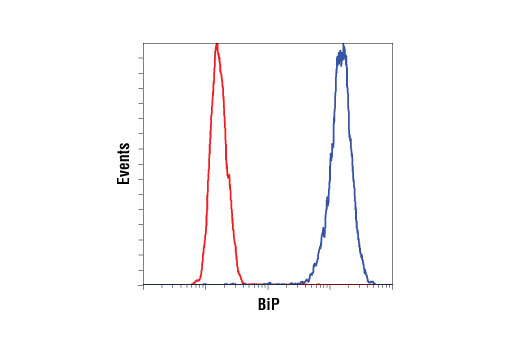
    Flow cytometric analysis of A204 cells using BiP (C50B12) Rabbit mAb (blue) compared to concentration-matched Rabbit (DA1E) mAb IgG XP® Isotype Control #3900 (red). Anti-rabbit IgG (H+L), F(ab')2 Fragment (Alexa Fluor® 488 Conjugate) #4412 was used as a secondary antibody.

    声明 :本官网所有报价均为常温或者蓝冰运输价格,如有产品需要干冰运输,需另外加收干冰运输费。
    货号:
    89947T
    一键复制
    询价
    供应商现货
    1Kit
    选择数量
    当前规格1件起购 
    配送至
    预计5-6个工作日送达,快递: 免运费,若需干冰额外收费