


 下载产品说明书 下载SDS
 用小程序,查商品更便捷


 收藏
收藏
 对比
对比 咨询
咨询
参考图片
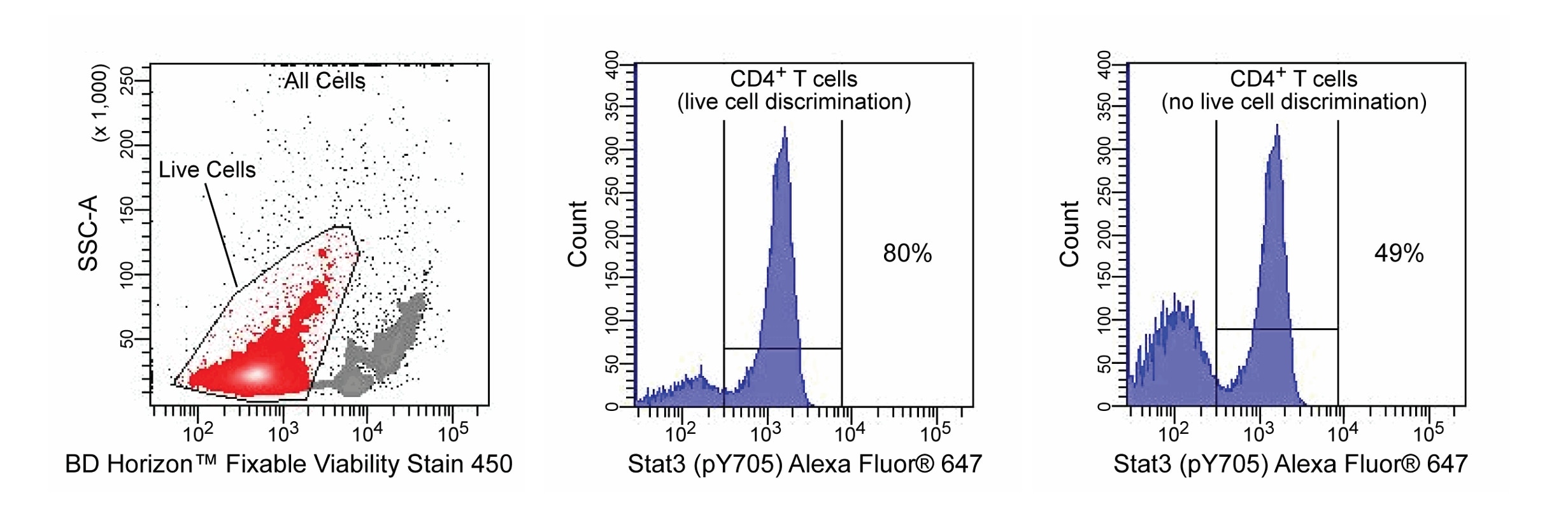
Multicolor flow cytometric analysis of phosphorylated STAT3 expression by “viable” activated human peripheral blood mononuclear cells (PBMC). PBMC were cultured for 48 hours in complete tissue culture medium and then frozen and stored (-80°C) for ten days. The cells were thawed and treated with recombinant human IL-6 (100 ng/ml; Cat. No. 550071) for 15 minutes with BD Horizon™ Fixable Viability Stain 450 (Cat. No. 562247) added for the last 7 minutes of activation. Cells were then fixed with BD Cytofix™ Fixation Buffer (Cat. No. 554655) and permeabilized with BD Phosflow™ Perm Buffer III (Cat. No. 558050) according to the standard Phosflow protocol. Cells were stained with PE Mouse Anti-Human CD3 (Cat. No. 555333), PerCP-Cy™5.5 Mouse Anti-Human CD4 (Cat. No. 552838) and BD Phosflow™ Alexa Fluor® 647 Mouse Anti-Stat3 (pY705) (Cat. No. 557815) antibodies. The dual parameter flow cytometric dot plot (Left Panel) shows the incorporated levels of FVS450 versus side scattered light signals expressed by the PBMC. Flow cytometric histograms show the levels of Stat3 (pY705) expressed by live cell-discriminated (ie, gated events with low level FVS450 incorporation; Middle Panel) and total events (including cells with both low and high levels of FVS450; Right Panel). The CD4+CD3+ T lymphocytes were derived from gated events with the forward and side light-scatter characteristics of intact lymphocytes. Flow cytometry was performed using a BD LSRFortessa™ Flow Cytometer System. FVS450 was also tested in mouse (data not shown).
Multicolor flow cytometric analysis of phosphorylated STAT3 expression by \"viable\" activated human peripheral blood mononuclear cells (PBMC). PBMC were cultured for 48 hours in complete tissue culture medium and then frozen and stored (-80°C) for ten days. The cells were thawed and treated with recombinant human IL-6 (100 ng/ml; Cat. No. 550071) for 15 minutes with BD Horizon™ Fixable Viability Stain 450 (Cat. No. 562247) added for the last 7 minutes of activation. Cells were then fixed with BD Cytofix™ Fixation Buffer (Cat. No. 554655) and permeabilized with BD Phosflow™ Perm Buffer III (Cat. No. 558050) according to the standard Phosflow protocol. Cells were stained with PE Mouse Anti-Human CD3 (Cat. No. 555333), PerCP-Cy™5.5 Mouse Anti-Human CD4 (Cat. No. 552838) and BD Phosflow™ Alexa Fluor® 647 Mouse Anti-Stat3 (pY705) (Cat. No. 557815) antibodies. The dual parameter flow cytometric dot plot (Left Panel) shows the incorporated levels of FVS450 versus side scattered light signals expressed by the PBMC. Flow cytometric histograms show the levels of Stat3 (pY705) expressed by live cell-discriminated (ie, gated events with low level FVS450 incorporation; Middle Panel) and total events (including cells with both low and high levels of FVS450; Right Panel). The CD4+CD3+ T lymphocytes were derived from gated events with the forward and side light-scatter characteristics of intact lymphocytes. Flow cytometry was performed using a BD LSRFortessa™ Flow Cytometer System. FVS450 was also tested in mouse (data not shown).

 危险品化学品经营许可证(不带存储) 许可证编号:沪(杨)应急管危经许[2022]202944(QY)
  营业执照(三证合一)