


BD Horizon™ PE-CF594 Mouse Anti-Human FoxP3


 下载产品说明书
 用小程序,查商品更便捷


 收藏
收藏
 对比
对比 咨询
咨询

参考图片
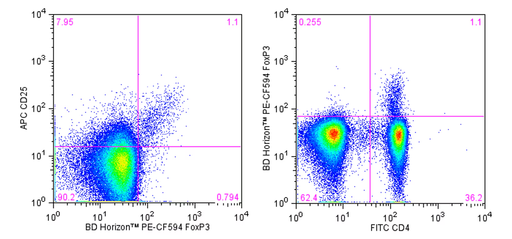
Multicolor flow cytometric analysis of FoxP3 expressed in human lymphocytes. Human peripheral blood mononuclear cells (PBMC) were stained with FITC Mouse Anti-Human CD4 (Cat. No. 555346/561842/561005) and APC anti-Human CD25 (Cat. No. 555434/560987) antibodies. The cells were then fixed and permeabilized (see Recommended Assay Procedure) and stained with BD Horizon™ PE-CF594 Mouse Anti-Human FoxP3 antibody (Cat No. 562421). Two-color flow cytometric dot plots show the correlated expression of either CD25 (Left Panel) or CD4 (Right Panel) versus FoxP3 derived from gated events with the light scattering characteristics of intact lymphocytes. Flow cytometry was performed using a BD™ LSR II Flow Cytometry System.
Multicolor flow cytometric analysis of FoxP3 expressed in human lymphocytes. Human peripheral blood mononuclear cells (PBMC) were stained with FITC Mouse Anti-Human CD4 (Cat. No. 555346/561842/561005) and APC anti-Human CD25 (Cat. No. 555434/560987) antibodies. The cells were then fixed and permeabilized (see Recommended Assay Procedure) and stained with BD Horizon™ PE-CF594 Mouse Anti-Human FoxP3 antibody (Cat No. 562421). Two-color flow cytometric dot plots show the correlated expression of either CD25 (Left Panel) or CD4 (Right Panel) versus FoxP3 derived from gated events with the light scattering characteristics of intact lymphocytes. Flow cytometry was performed using a BD™ LSR II Flow Cytometry System.

 危险品化学品经营许可证(不带存储) 许可证编号:沪(杨)应急管危经许[2022]202944(QY)
  营业执照(三证合一)