


 用小程序,查商品更便捷


ICC
1:500FC
1:2000IF
1:500
N/A
12 months from date of receipt / reconstitution, 2 to 8 °C as supplied
参考图片
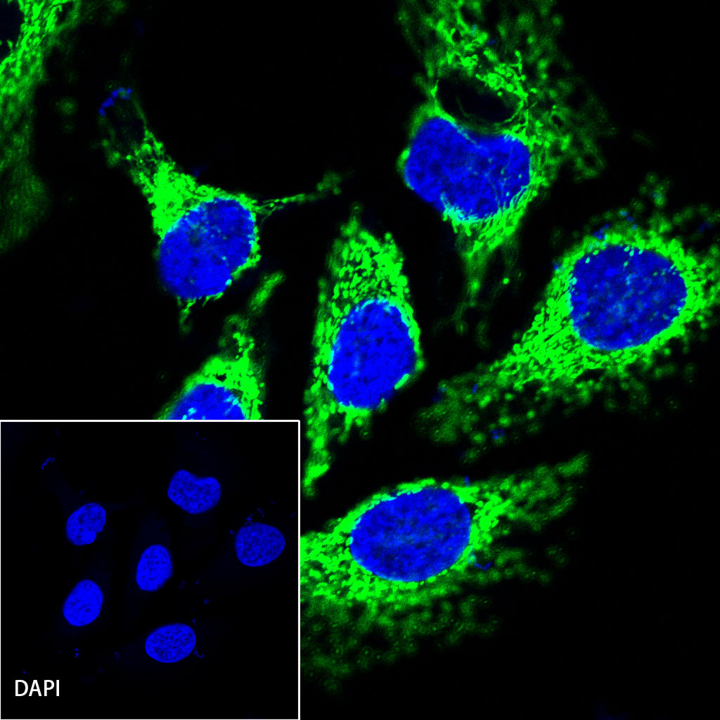
ICC shows positive staining in HeLa cells. Anti-HSP60 antibody (S0B0002) was used at 1/500 dilution (Green) and incubated overnight at 4°C. Goat anti-Rabbit IgG(H+L) (Alexa Fluor® 488 Conjugate) was used as secondary antibody at 1/500 dilution. The cells were fixed with 4% PFA and permeabilized with 0.1% PBS-Triton X-100. Nuclei were counterstained with DAPI (Blue).
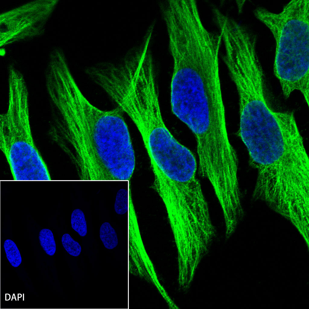
ICC shows positive staining in HeLa cells. Anti-α-tubulin antibody (S0B0043) was used at 1/100 dilution (Green) and incubated overnight at 4°C. Goat anti-Rabbit IgG(H+L) (Alexa Fluor® 488 Conjugate) was used as secondary antibody at 1/500 dilution. The cells were fixed with 4% PFA and permeabilized with 0.1% PBS-Triton X-100. Nuclei were counterstained with DAPI (Blue).
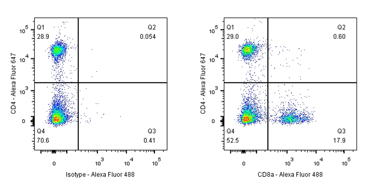
Flow cytometric analysis of mouse splenocytes labeling CD8a antibody (S0B0034) at 1/50 dilution (1 μg) (Right) compared with a Rabbit monoclonal IgG isotype control (Left). Goat anti-Rabbit IgG(H+L) (Alexa Fluor® 488 Conjugate) was used as the secondary antibody at 1/2000 dilution.
Cells were surface stained with CD4-Alexa Fluor® 647, then stained with rabbit IgG (Left) / anti-CD8a (Right) separately. CD8a and CD4 are mutually exclusive expressed in mouse splenocytes. Gated on total viable cells.
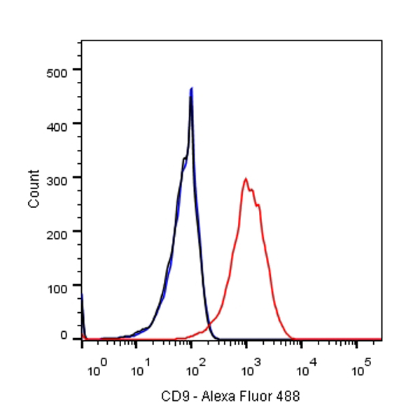
Flow cytometric analysis of HCT 116 cells labelling CD9 antibody (S0B0027) at 1/500 (0.1 μg) dilution/ (red) compared with a Rabbit monoclonal IgG (Black) isotype control and an unlabelled control (cells without incubation with primary antibody and secondary antibody) (Blue). Goat anti-Rabbit IgG(H+L) (Alexa Fluor® 488 Conjugate) was used as the secondary antibody at 1/2000 dilution.
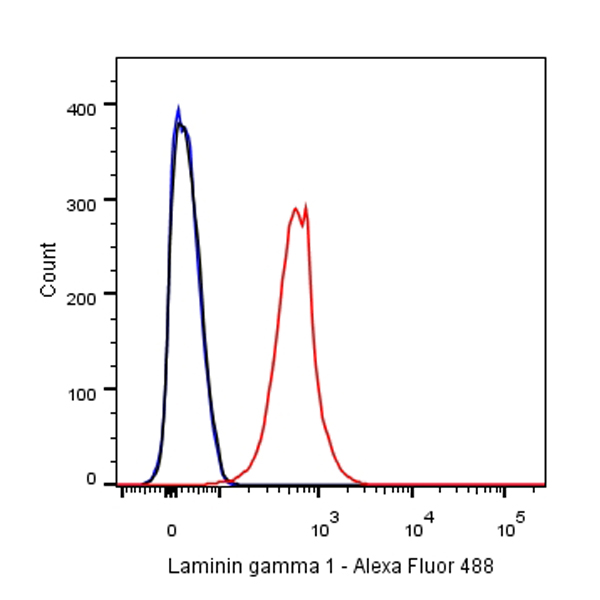
Flow cytometric analysis of A431 cells labelling Laminin gamma 1 antibody (S0B0069) at 1/500 (0.1 μg) dilution/ (red) compared with a Rabbit monoclonal IgG (Black) isotype control and an unlabelled control (cells without incubation with primary antibody and secondary antibody) (Blue). Goat anti-Rabbit IgG(H+L) (Alexa Fluor® 488 Conjugate) was used as the secondary antibody at 1/2000 dilution.
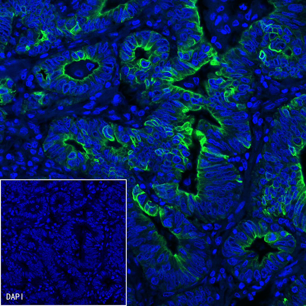
IF shows positive staining in paraffin-embedded human colon cancer. Anti-CK-LMW antibody was used at 1/500 dilution (Green) and incubated overnight at 4°C. Goat anti-Rabbit IgG(H+L) (Alexa Fluor® 488 Conjugate) (S0B4004) was used as secondary antibody at 1/500 dilution. Counterstained with DAPI (Blue). Heat mediated antigen retrieval with EDTA buffer pH9.0 was performed before commencing with IF staining protocol.
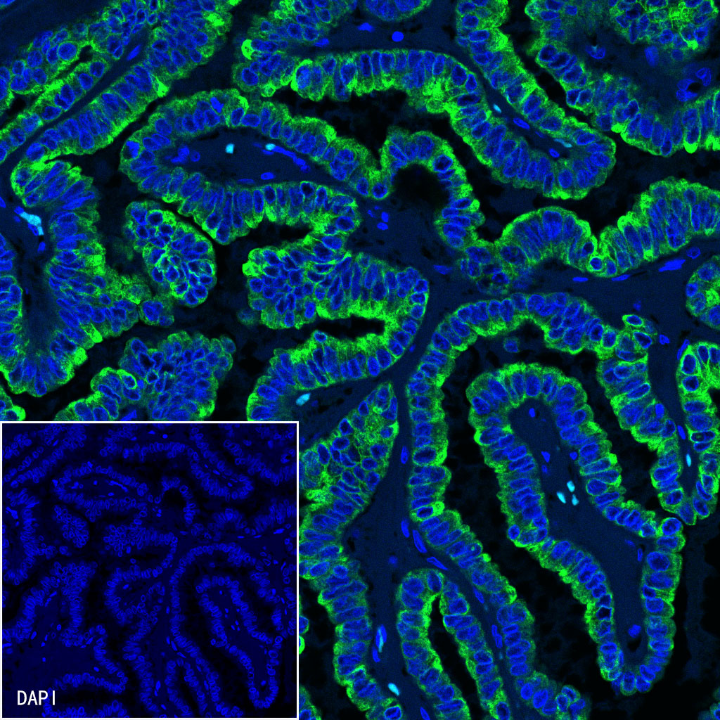
IF shows positive staining in paraffin-embedded human thyroid cancer. Anti-CK-LMW antibody was used at 1/500 dilution (Green) and incubated overnight at 4°C. Goat anti-Rabbit IgG(H+L) (Alexa Fluor® 488 Conjugate) (S0B4004) was used as secondary antibody at 1/500 dilution. Counterstained with DAPI (Blue). Heat mediated antigen retrieval with EDTA buffer pH9.0 was performed before commencing with IF staining protocol.
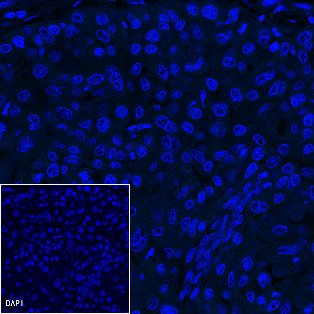
Negative control: IF shows negative staining in paraffin-embedded human keratinized oral squamous cell carcinoma. Anti-CK-LMW antibody was used at 1/500 dilution and incubated overnight at 4°C. Goat anti-Rabbit IgG(H+L) (Alexa Fluor® 488 Conjugate) (S0B4004) was used as secondary antibody at 1/500 dilution. Counterstained with DAPI (Blue). Heat mediated antigen retrieval with EDTA buffer pH9.0 was performed before commencing with IF staining protocol.

 危险品化学品经营许可证(不带存储) 许可证编号:沪(杨)应急管危经许[2022]202944(QY)
  营业执照(三证合一)