


 用小程序,查商品更便捷


ICC
1:500IF
1:500
12 months from date of receipt / reconstitution, 2 to 8 °C as supplied
参考图片
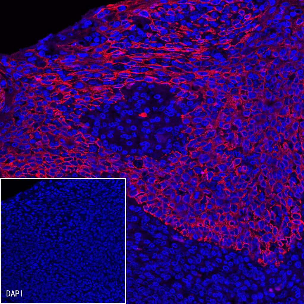
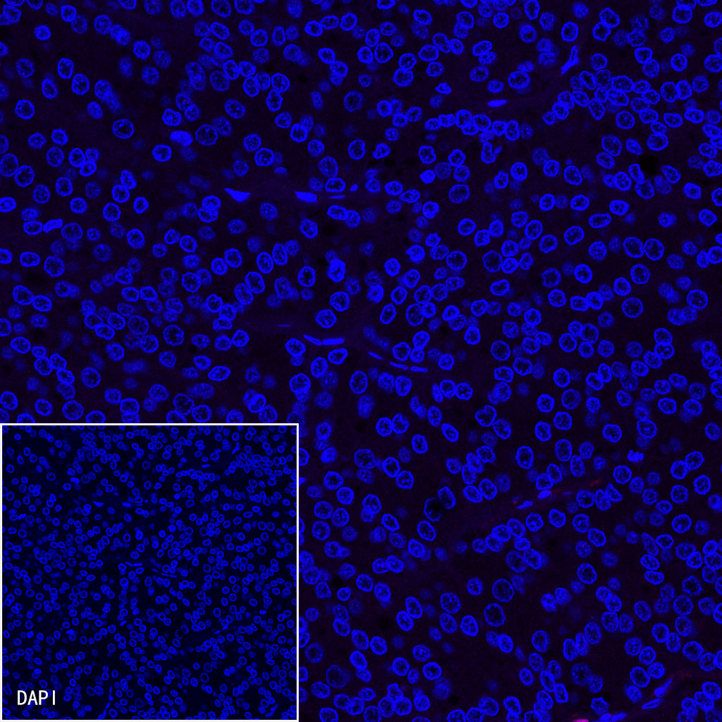
ICC shows positive staining in HeLa cells. Anti-HSP60 antibody (S0B0002) was used at 1/500 dilution (Red) and incubated overnight at 4°C. Goat anti-Rabbit IgG (H+L) (Alexa Fluor® 594 Conjugate) was used as secondary antibody at 1/500 dilution. The cells were fixed with 4% PFA and permeabilized with 0.1% PBS-Triton X-100. Nuclei were counterstained with DAPI (Blue).
ICC shows positive staining in MCF7 cells. Anti-HSP60 antibody (S0B0002) was used at 1/500 dilution (Red) and incubated overnight at 4°C. Goat anti-Rabbit IgG (H+L) (Alexa Fluor® 594 Conjugate) was used as secondary antibody at 1/500 dilution. The cells were fixed with 4% PFA and permeabilized with 0.1% PBS-Triton X-100. Nuclei were counterstained with DAPI (Blue).
ICC shows positive staining in HeLa cells. Anti-α-tubulin antibody (S0B0043) was used at 1/500 dilution (Red) and incubated overnight at 4°C. Goat anti-Rabbit IgG (H+L) (Alexa Fluor® 594 Conjugate) was used as secondary antibody at 1/500 dilution. The cells were fixed with 4% PFA and permeabilized with 0.1% PBS-Triton X-100. Nuclei were counterstained with DAPI (Blue).
ICC shows positive staining in MCF7 cells. Anti-α-tubulin antibody (S0B0043) was used at 1/500 dilution (Red) and incubated overnight at 4°C. Goat anti-Rabbit IgG (H+L) (Alexa Fluor® 594 Conjugate) was used as secondary antibody at 1/500 dilution. The cells were fixed with 4% PFA and permeabilized with 0.1% PBS-Triton X-100. Nuclei were counterstained with DAPI (Blue).

 危险品化学品经营许可证(不带存储) 许可证编号:沪(杨)应急管危经许[2022]202944(QY)
  营业执照(三证合一)