


 下载产品说明书
 用小程序,查商品更便捷


 收藏
收藏
 对比
对比 咨询
咨询
Product Usage Information
Directions for Use: DAPI is supplied as a lyophilized powder. For a 20 mg/ml stock, reconstitute the 1 mg in 50 µl deionized water or dimethylformamide. Please follow CST’s recommended IF and Flow protocols. For both applications, following secondary detection:Immunofluorescence: Counterstain with DAPI as the final step in your staining procedure. Rinse samples twice in PBS for five min each. Dilute DAPI stock solution to a concentration between 1 - 0.1 µg/ml in PBS and incubate for 5 min at room temperature in the dark. Rinse samples once in PBS and then prepare for imaging. Examine immediately using appropriate excitation wavelength.Alternatively, dilute DAPI stock solution to a concentration between 1 - 0.1 µg/ml in mounting media, apply to cells, and prepare for imaging. Examine immediately using appropriate excitation wavelength.Flow Cytometry: Rinse samples once in Incubation Buffer. Dilute DAPI stock solution to a concentration between 1.60-0.400 µg/ml in PBS and incubate for 15 min at room temperature in the dark before analyzing cells on flow cytometer.
参考图片
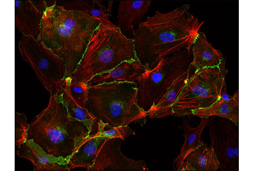
Immunofluorescent analysis of HUVE cells using VE-Cadherin (D87F2) XP® Rabbit mAb #2500 (green) and DAPI (blue). Actin filaments were labeled with DY-554 phalloidin (red).免疫荧光分析HUVE细胞,所用抗体为VE-Cadherin (D87F2) XP™ Rabbit mAb #2500 (绿色) and DAPI (蓝色).肌动蛋白丝采用DY-554 phalloidin (红色)标记。
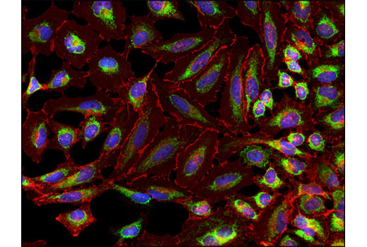
Immunofluorescent analysis of HeLa cells using COX IV (3E11) Rabbit mAb #4850 (green), β-Catenin (L54E2) Mouse mAb (IF Preferred) #2677 (red) and DAPI (blue).免疫荧光分析HeLa细胞,所用抗体为g COX IV (3E11) Rabbit mAb #4850 (绿色), β-Catenin (L54E2) Mouse mAb (IF Preferred) #2677 (红色) 和 DAPI (蓝色)。
Flow cytometric analysis of Jurkat cells using Ki-67 (D3B5) Rabbit mAb (Alexa Fluor® 647 Conjugate) #12075 and DAPI (DNA content).

 危险品化学品经营许可证(不带存储) 许可证编号:沪(杨)应急管危经许[2022]202944(QY)
  营业执照(三证合一)