


 下载产品说明书 下载SDS
 用小程序,查商品更便捷


 收藏
收藏
 对比
对比 咨询
咨询
Product Usage Information
As a last step for fluorescent assays, add enough neat (undiluted) PI/RNase Staining Solution to submerge cells or samples. Incubate for 15 minutes at room temperature, protected from light before analysis.
参考图片
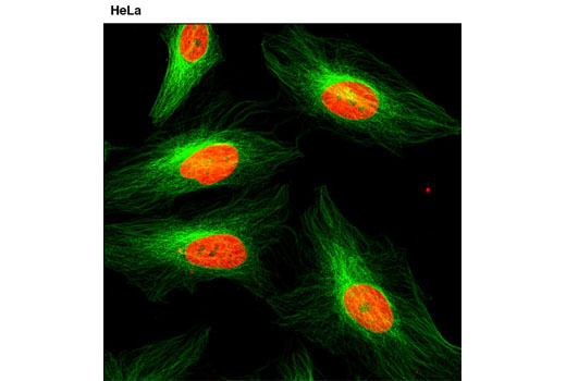
Confocal immunofluorescent analysis of HeLa cells using α-Tubulin (DM1A) Mouse mAb #3873 (green). Red = Propidium Iodide (PI)/RNase Staining Solution.激光共聚焦免疫荧光分析HeLa细胞,所用抗体为α-Tubulin (DM1A) Mouse mAb #3873 (绿色). 红色为碘化丙啶(PI )/核糖核酸酶染色试剂。
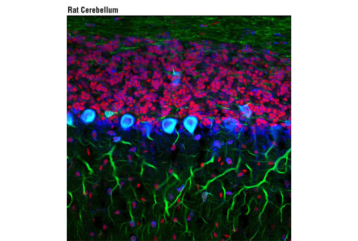
Confocal immunofluorescent analysis of rat cerebellum using S6 Ribosomal Protein (54D2) Mouse mAb (Alexa Fluor® 647 Conjugate) #5548 (blue pseudocolor) and β3-Tubulin (D71G9) XP® Rabbit mAb #5568 (green). Red = Propidium Iodide (PI)/RNase Staining Solution.激光共聚焦免疫荧光分析大鼠小脑细胞,所用抗体为S6 Ribosomal Protein (54D2) Mouse mAb (Alexa Fluor ® 647 Conjugate) #5548 (blue pseudocolor) 和 β3-Tubulin (D71G9) XP ® Rabbit mAb #5568 (绿色),红色为碘化丙啶(PI )/核糖核酸酶染色试剂(DNA)
Flow cytometric analysis of untreated Jurkat cells, using Phospho-Histone H3 (Ser10) Antibody (Alexa Fluor® 488 Conjugate) #9708 and Propidium Iodide (PI)/RNase Staining Solution (DNA content).流式细胞术分析未处理的Jurkat 细胞,所用抗体为Phospho-Histone H3 (Ser10) Antibody (Alexa Fluor® 488 Conjugate) #9708 和 碘化丙啶 (PI)/RNase Staining Solution (DNA content).

 危险品化学品经营许可证(不带存储) 许可证编号:沪(杨)应急管危经许[2022]202944(QY)
  营业执照(三证合一)