


 用小程序,查商品更便捷


Product Usage Information
The optimal dilution of the anti-species antibody should be determined for each primary antibody by titration. However, a final dilution of 1:500–1:2000 should yield acceptable results for immunofluorescent and flow cytometry assays.
Specificity/Sensitivity
Species Reactivity:
Rabbit
This product has been optimized for use as a secondary antibody in immunofluorescent applications. Fluorescent anti-species IgG conjugates are ideal for flow cytometry and immunofluorescence. Cell Signaling Technology’s strict quality control procedures assure that each conjugate provides optimal specificity and fluorescence.
参考图片
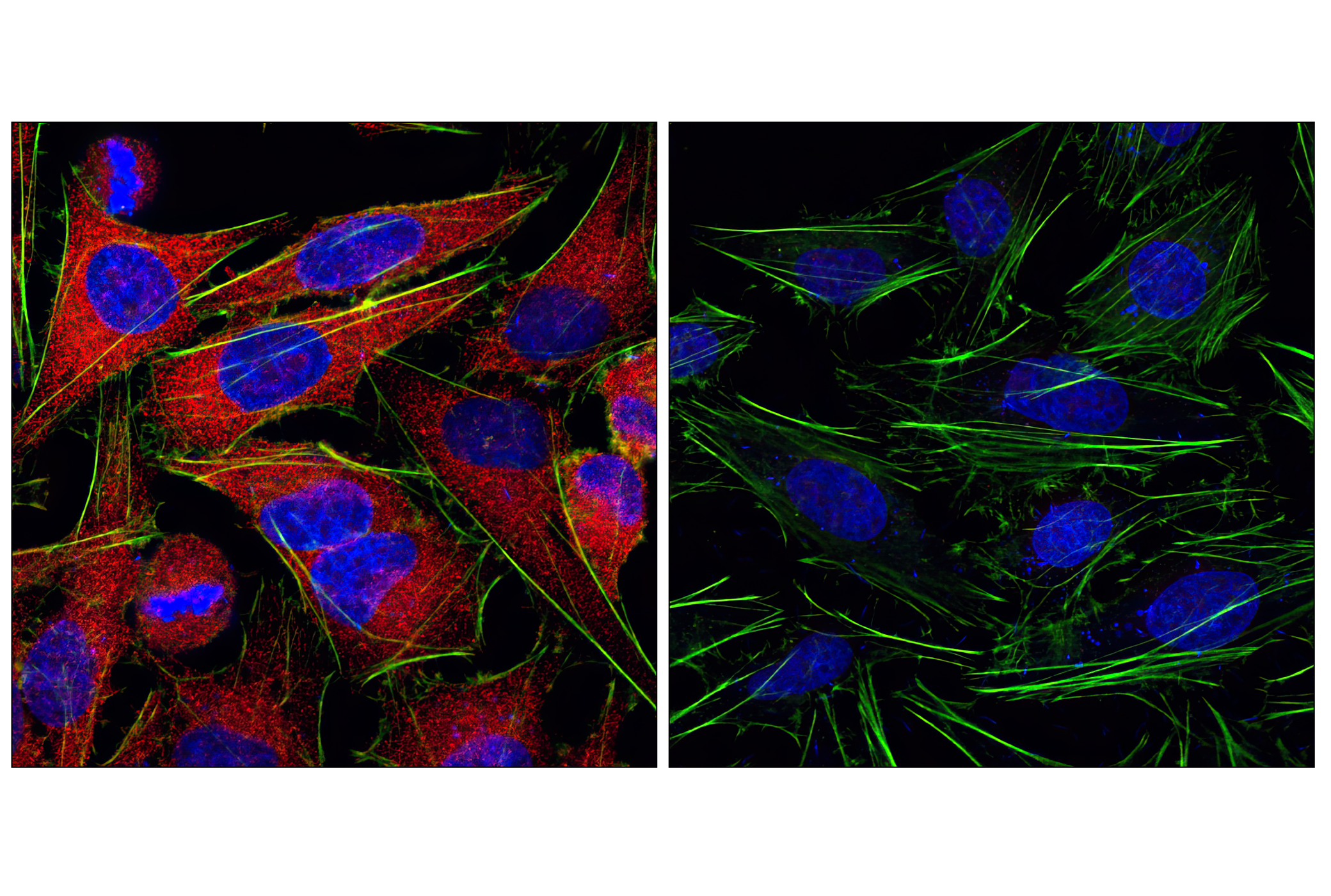
Confocal immunofluorescent analysis of HeLa cells labeled with MEK1/2 (47E6) Rabbit mAb #9126 detected with Anti-Rabbit IgG (H+L), F(ab')2 Fragment (Alexa Fluor® 555 Conjugate) (red, left) compared to an isotype control (right). Actin filaments have been labeled with fluorescein phalloidin (green). Blue pseudocolor = DRAQ5® #4084 (fluorescent DNA dye).激光共聚焦免疫荧光分析HeLa细胞,所用抗体为MEK1/2 (47E6) Rabbit mAb #9126检测抗体为anti- Rabbit IgG (H+L), F(ab’)2 Fragment (Alexa Fluor ® 555 Conjugate) (红色,上图),与同型对照(下图)作比较。肌动蛋白丝采用fluorescein phalloidin (绿色),蓝色伪彩为荧光DNA染料,信息为DRAQ5 ® #4084。
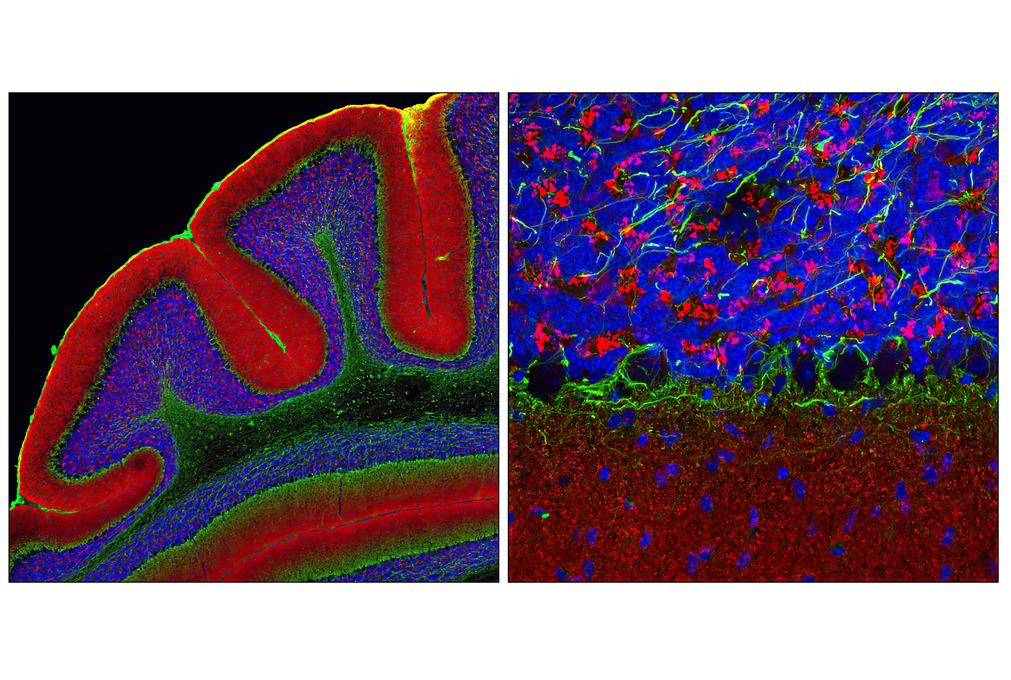
Confocal immunofluorescent analysis of mouse cerebellum using α-Synuclein Antibody (IF Preferred) #2628 detected with Anti-Rabbit IgG (H+L), F(ab')2 Fragment (Alexa Fluor® 555 Conjugate) (red) and Neurofilament-L (DA2) Mouse mAb #2835 detected with Anti-Mouse IgG (H+L), F(ab')2 Fragment (Alexa Fluor® 488 Conjugate) #4408 (green). Blue pseudocolor = DRAQ5® #4084 (fluorescent DNA dye).激光共聚焦免疫荧光分析鼠小脑细胞,所用抗体为α-Synuclein Antibody (IF Preferred) #2628检测抗体为anti- Rabbit IgG (H+L), F(ab’)2 Fragment (Alexa Fluor ® 555 Conjugate) (红色)和Neurofilament-L (DA2) Mouse mAb #2835,检测抗体为Anti-Mouse IgG (H+L), F(ab’)2 Fragment (Alexa Fluor ® 488 Conjugate) #4408 (绿色) 。蓝色伪彩为荧光DNA染料,信息为DRAQ5 ® #4084。
High content analysis of A549 cells exposed to varying concentrations of LY294002 #9901 for 3 hrs, followed by 100 ng/mL EGF for 20 minutes. With increasing concentrations of LY294002, a significant decrease (~5 fold) in phospho-S6 Ribosomal Protein (Ser235/236) signal as compared to the uninhibited control was observed. When using phospho-S6 as a measurement, the IC50 of this compound was 3.06 μM. Data were generated on the Acumen HCS platform using Anti-Rabbit IgG (H+L), F(ab')2 Fragment (Alexa Fluor® 555 Conjugate).图 高通量分析A549细胞,该细胞暴露于不同浓度的LY294002 #9901 3个小时。然后用100ng/mlde EGF 处理20分钟。与未抑制组相比较,随着LY294002 #9901浓度的增加phospho-S6 Ribosomal Protein (Ser235/236)信号显著降低(约5倍)。当phospho-S6 Ribosomal Protein 检测时,IC50为3.06mM。数据时以Acumen ® HCS为平台生成的,所用抗体为Anti-Rabbit IgG (H+L), F(ab’)2 Fragment (Alexa Fluor ® 555 Conjugate)。

 危险品化学品经营许可证(不带存储) 许可证编号:沪(杨)应急管危经许[2022]202944(QY)
  营业执照(三证合一)