


 用小程序,查商品更便捷


Product Usage Information
Important! This control antibody must be diluted to the same concentration (not dilution) as the specific antibody used in analysis. Higher background fluorescence may result if excessive amounts of isotype control are used. For protocol details, please reference the product page for the specific antibody used in analysis.
Specificity/Sensitivity
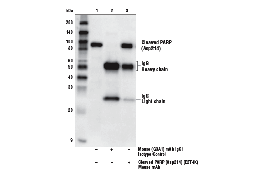
参考图片
Confocal immunofluorescent analysis of HT-29 cells using α-Tubulin (DM1A) Mouse mAb #3873 (green, left) compared to concentration matched Mouse (G3A1) mAb IgG1 Isotype Control (green, right). Blue pseudocolor = DRAQ5® #4084 (fluorescent DNA dye).
Confocal immunofluorescent analysis of normal rat cerebellum using Neurofilament H (RMdO 20) Mouse mAb #2836 (green, left) compared to concentration matched Mouse (G3A1) mAb IgG1 Isotype Control (green, right). Blue pseudocolor = DRAQ5® #4084 (fluorescent DNA dye).激光共聚焦荧光法检测正常大鼠小脑,所用检测抗体为Neurofilament H (RMdO 20) Mouse mAb #2836 (绿色,上图) 与浓度匹配的 Mouse (G3A1) mAb IgG1 Isotype Control (绿色,下图)作比较。蓝色伪彩为DNA荧光染料(产品信息DRAQ5®#4084 )。
Chromatin immunoprecipitations were performed using digested chromatin from HeLa cells and the indicated antibodies. Purified DNA was analyzed by quantitative real-time PCR, using SimpleChIP® Human GAPDH Exon 1 Primers #5516, SimpleChIP® Human RPL30 Exon 3 Primers #7014, and SimpleChIP® Human MYT-1 Exon 1 Primers #4493. The relative abundance of each DNA sequence enriched by Mouse (G3A1) mAb IgG1 Isotype Control (red) is compared to the amount of the same DNA sequence enriched by the histone H3-specific immunoprecipitations (blue).采用HeLa细胞已消化的染色质和已知抗体进行染色质免疫沉淀。实时定量PCR检测纯化DNA,所用引物分别为SimpleChIP ® Human GAPDH Exon 1 Primers #5516, SimpleChIP ® Human RPL30 Exon 3 Primers #7014, 和SimpleChIP ® Human MYT-1 Exon 1 Primers #4493. 每一条DNA序列的相对含量,所用抗体为Mouse (G3A1) mAb IgG1 同型对照(红色) 与同一条DNA序列的含量做比较,后者通过为组蛋白H3特异的免疫沉淀检测(蓝色)。
Flow cytometric analysis of MCF7 cells using Pan-Keratin (C11) Mouse mAb #4545 (blue) compared to concentration-matched Mouse (G3A1) mAb IgG1 Isotype Control (red).流式细胞术分析MCF7细胞,所用检测抗体为Pan-Keratin (C11) Mouse mAb #4545 (蓝色)与浓度匹配的 Mouse (G3A1) mAb IgG1 同型对照(红色)作比较。
Flow cytometric analysis of Jurkat cells, U0126-treated (blue) or TPA-treated (green), using Phospho-p44/42 MAPK (Erk1/2) (Thr202/Tyr204) (E10) Mouse mAb #9106 compared to concentration-matched Mouse (G3A1) mAb IgG1 Isotype Control (red).流式细胞术检测Jurkat细胞,蓝色为U0126处理组,绿色为TPA处理组。所用检测抗体为Phospho-p44/42 MAPK (Erk1/2) (Thr202/Tyr204) (E10) Mouse mAb #9106 与浓度匹配的 Mouse (G3A1) mAb IgG1 同型对照(红色)作比较.
Immunohistochemical analysis of paraffin-embedded human seminoma using Nanog (1E6C4) Mouse mAb #4893 (left) or Mouse (G3A1) mAb IgG1 Isotype Control (right).免疫组化分析人类精原细胞瘤 。所用检测抗体为Nanog (1E6C4) Mouse mAb #4893 (上图)或Mouse (G3A1) mAb IgG1 同型对照(右图)。

 危险品化学品经营许可证(不带存储) 许可证编号:沪(杨)应急管危经许[2022]202944(QY)
  营业执照(三证合一)