


 用小程序,查商品更便捷


Product Usage Information
Important! This control antibody must be diluted to the same concentration (not dilution) as the specific antibody used in analysis. Higher background fluorescence may result if excessive amounts of isotype control are used. For protocol details, please reference the product page for the specific antibody used in analysis.
Specificity/Sensitivity
参考图片
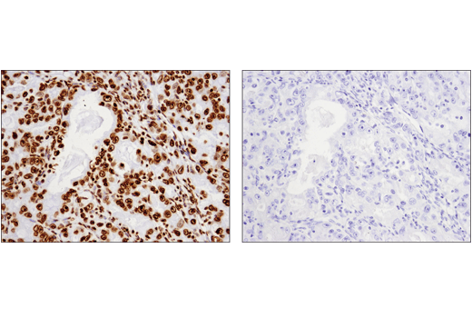
Immunohistochemical analysis of paraffin-embedded human ovarian carcinoma using Histone H3 (1B1B2) Mouse mAb #14269 (left) compared to concentration-matched Mouse (E1D5H) mAb IgG3 Isotype Control (right).
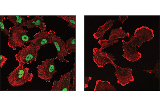
Confocal immunofluorescent analysis of SNB-19 cells using Histone H3 (1B1B2) Mouse mAb #14269 (left, green) compared to concentration-matched Mouse (E1D5H) mAb IgG3 Isotype Control (right, green). Cells were counterstained with β-Actin (13E5) Rabbit mAb #4970 (red).
Flow cytometric analysis of K-562 cells using Histone H3 (1B1B2) Mouse mAb #14269 (solid line) compared to concentration-matched Mouse (E1D5H) mAb IgG3 Isotype Control (dashed line). Anti-mouse IgG (H+L), F(ab')2 Fragment (Alexa Fluor® 488 Conjugate) #4408 was used as a secondary antibody.
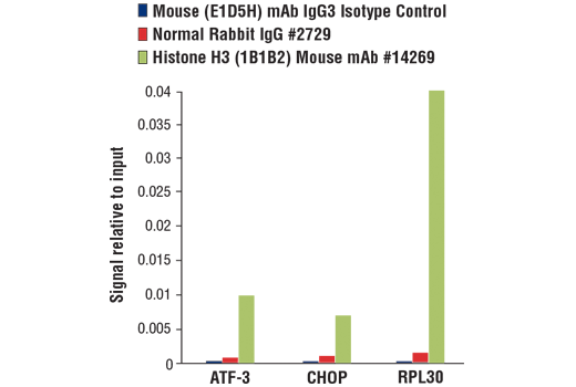
Chromatin immunoprecipitations were performed with cross-linked chromatin from 4 x 10⁶ MEF cells treated with tunicamycin (2 μg/ml, 10 hr) and either Mouse (E1D5H) mAb IgG3 Isotype Control, Normal Rabbit IgG #2729, or Histone H3 (1B1B2) Mouse mAb #14269 using SimpleChIP® Plus Enzymatic Chromatin IP Kit (Magnetic Beads) #9005. The enriched DNA was quantified by real-time PCR using SimpleChIP® Mouse ATF-3 Intron 1 Primers #13059, mouse CHOP promoter primers, and SimpleChIP® Mouse RPL30 Intron 2 Primers #7015. The amount of immunoprecipitated DNA in each sample is represented as signal relative to the total amount of input chromatin, which is equivalent to one.

 危险品化学品经营许可证(不带存储) 许可证编号:沪(杨)应急管危经许[2022]202944(QY)
  营业执照(三证合一)