


 下载产品说明书
 用小程序,查商品更便捷


 收藏
收藏
 对比
对比 咨询
咨询
Specificity/Sensitivity
参考图片
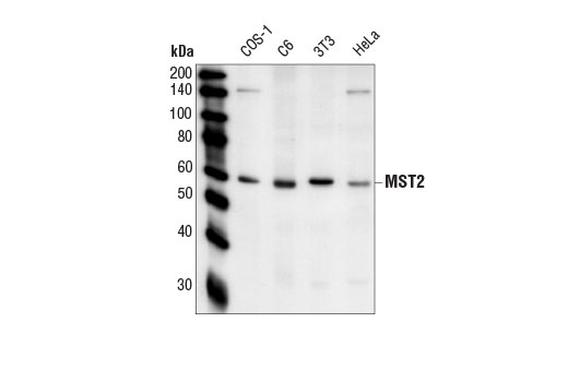
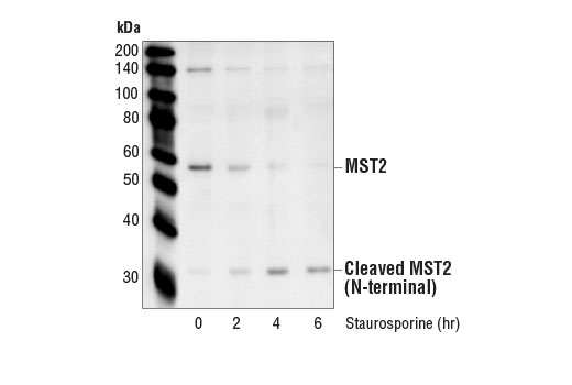
Western blot analysis of extracts from various cell lines using Mst2 Antibody #3952.
Western blot analysis of extracts from various cell lines using Sav1 Antibody #3507.
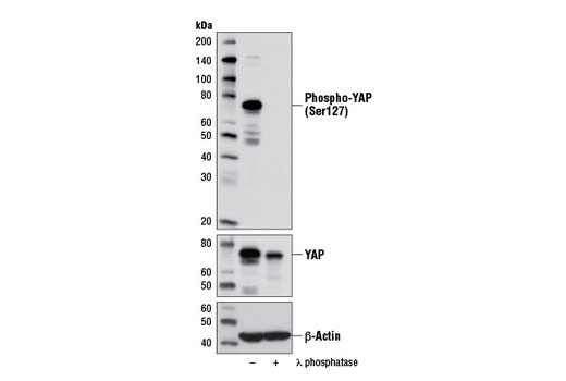
Western blot analysis of extracts from PANC-1 cells, untreated (-) or λ-phosphatase-treated (+), using Phospho-YAP (Ser127) (D9W2I) Rabbit mAb (upper), YAP Antibody #4912 (middle), and β-Actin (D6A8) Rabbit mAb #8457 (lower).
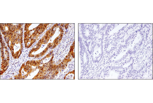
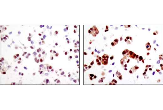
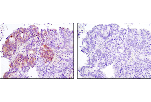
Immunohistochemical analysis of paraffin-embedded human colon adenocarcinoma, control (left) or λ-phosphatase treated (right), using Phospho-YAP (Ser127) (D9W2I) Rabbit mAb.
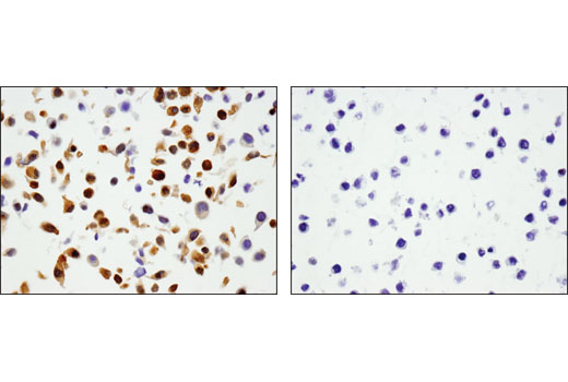
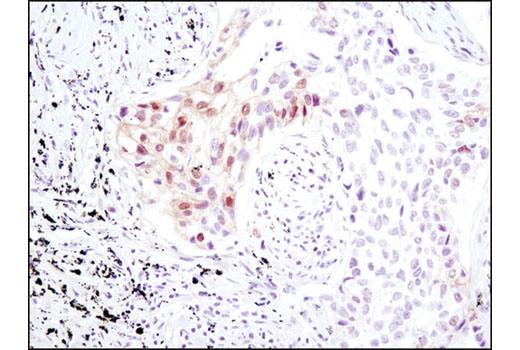
Immunohistochemical analysis of paraffin-embedded human non-small cell lung carcinoma using Phospho-YAP (Ser127) (D9W2I) Rabbit mAb in the presence of control peptide (left) or antigen-specific peptide (right).
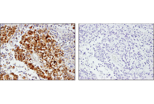
Immunohistochemical analysis of paraffin-embedded cell pellets, A-204 (left) and RL-7 (right), using Phospho-YAP (Ser127) (D9W2I) Rabbit mAb.
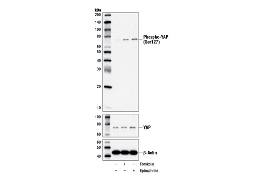
Western blot analysis of MDA-MB-231 cells, vehicle-treated (-) or treated with Forskolin #3828 (10 μM, 60 min; +) or epinephrine (10 μM, 60 min; +), using Phospho-YAP (Ser127) (D9W2I) Rabbit mAb (upper), YAP Antibody #4912 (middle), and β-Actin (D6A8) Rabbit mAb #8457 (lower).
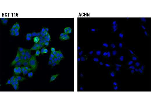
Confocal immunofluorescent analysis of HCT 116 (higher expressing, left) and ACHN (lower expressing, right) cells using SAV1 (D6M6X) Rabbit mAb (green). Blue pseudocolor = DRAQ5® #4084 (fluorescent DNA dye).
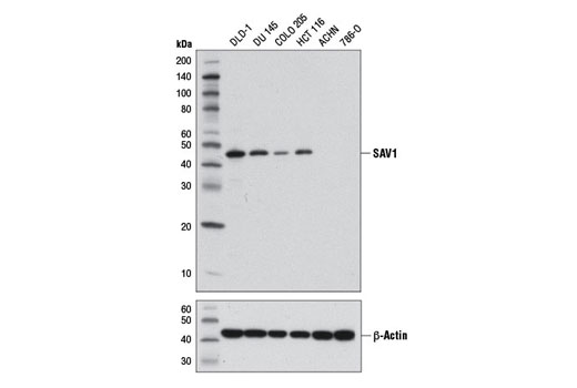
Western blot analysis of extracts from various cell lines using SAV1 (D6M6X) Rabbit mAb (upper) and β-Actin (D6A8) Rabbit mAb #8457 (lower).
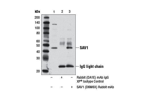
Immunoprecipitation of SAV1 protein from A-204 cell extracts using Rabbit (DA1E) mAb IgG XP® Isotype Control #3900 (lane 2) or SAV1 (D6M6X) Rabbit mAb (lane 3). Lane 1 is 10% input. Western blot analysis was performed using SAV1 (D6M6X) Rabbit mAb.
Immunohistochemical analysis of paraffin-embedded human lung carcinoma using Phospho-MOB1 (Thr35) (D2F10) Rabbit mAb.
Immunohistochemical analysis of paraffin-embedded human ovarian carcinoma control (left) or lambda phosphatase-treated (right) using Phospho-MOB1 (Thr35) (D2F10) Rabbit mAb.
Immunohistochemical analysis of paraffin-embedded MCF7 cell pellets, control (left) or H2O2-treated (right), using Phospho-MOB1 (Thr35) (D2F10) Rabbit mAb.
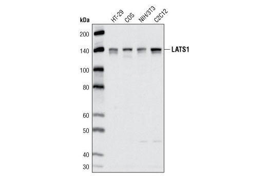
Western blot analysis of extracts from various cell lines using LATS1 (C66B5) Rabbit mAb.
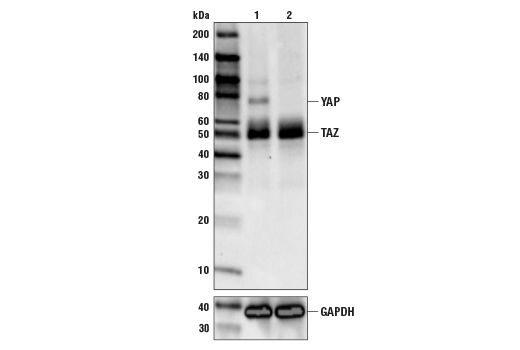
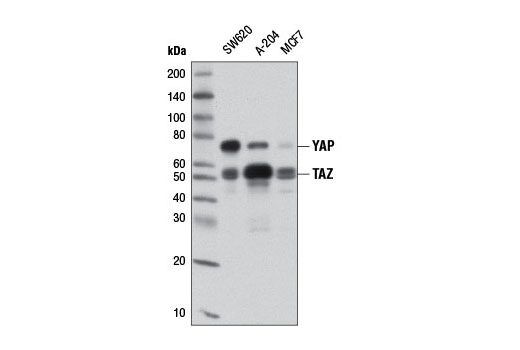
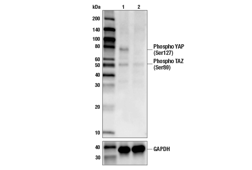
Western blot analysis of extracts from various cell lines using YAP/TAZ (D24E4) Rabbit mAb.
Immunohistochemical analysis of paraffin-embedded COS-7 cell pellets, transfected with YAP (left) or TAZ (right), using YAP/TAZ (D24E4) Rabbit mAb.
Immunohistochemical analysis of paraffin-embedded human lymphoma using YAP/TAZ (D24E4) Rabbit mAb.
Immunohistochemical analysis of paraffin-embedded human colon carcinoma using YAP/TAZ (D24E4) Rabbit mAb.
After the primary antibody is bound to the target protein, a complex with HRP-linked secondary antibody is formed. The LumiGLO* is added and emits light during enzyme catalyzed decomposition.
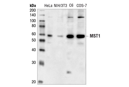
Western blot analysis of extracts from HeLa, NIH/3T3, C6 and COS cells, using Mst1 Antibody.
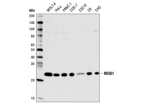
Western blot analysis of extracts from various cell lines using MOB1 Antibody.
Western blot analysis of extracts from HeLa cells, untreated (-) or okadaic acid-treated (+), using Phospho-LATS1 (Thr1079) (D57D3) Rabbit mAb (upper) or LATS1 (C66B5) Rabbit mAb #3477 (lower).
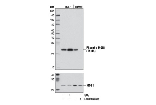
Western blot analysis of extracts from MCF7 cells, either untreated (-) or treated (+) with H2O2 (2.5 mM, 30 min) and Ramos cells, either untreated (-) or treated (+) with λ phosphatase, using Phospho-MOB1 (Thr35) (D2F10) Rabbit mAb (upper) and MOB1 Antibody #3863 (lower).
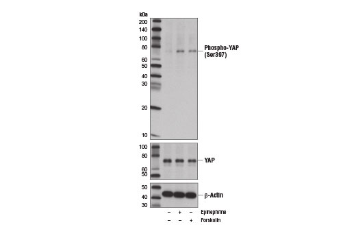
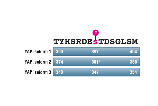
Western blot analysis of extracts from MDA-MB-231 cells, vehicle treated (-) or treated with epinephrine (10 μM, 60 min; +) or Forskolin #3828 (10 μm, 60 min; +), using Phospho-YAP (Ser397) (D1E7Y) Rabbit mAb (upper), YAP Antibody #4912 (middle), and β-Actin (D6A8) Rabbit mAb #8547 (lower). YAP protein isoform 1 Ser397 corresponds to Ser381 of YAP isoform 2, as reported by Zhao, B. et al. (2010) Genes Dev 24, 72-85 (9).
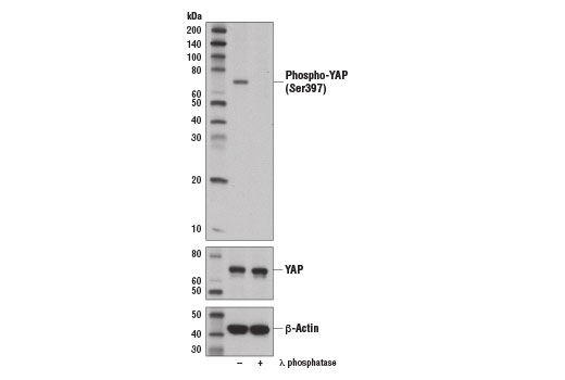
Western blot analysis of extracts from Hep G2 cells, untreated (-) or λ-phosphatase-treated (+), using Phospho-YAP (Ser397) (D1E7Y) Rabbit mAb (upper), YAP Antibody #4912 (middle), and β-Actin (D6A8) Rabbit mAb #8457 (lower). YAP protein isoform 1 Ser397 corresponds to Ser381 of YAP isoform 2, as reported by Zhao et al. (2010) Genes Dev 24, 72-85 (9).
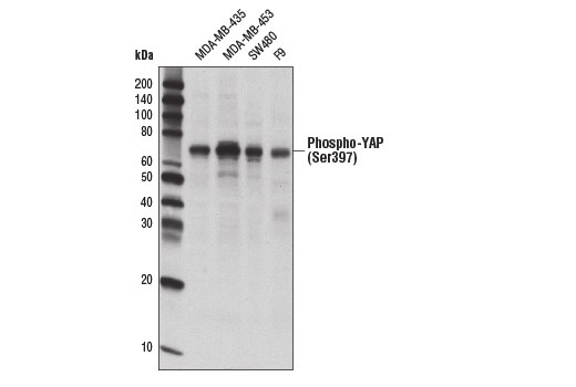
Western blot analysis of extracts from various cell lines using Phospho-YAP (Ser397) (D1E7Y) Rabbit mAb.
Western blot analysis of extracts from various cell lines using MOB1 (E1N9D) Rabbit mAb.
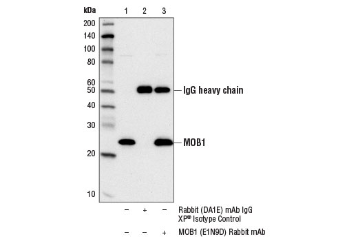
Immunoprecipitation of MOB1 protein from HeLa cell extracts using Rabbit (DA1E) mAb IgG XP® Isotype Control #3900 (lane 2) or MOB1 (E1N9D) Rabbit mAb (lane 3). Lane 1 is 10% input. Western blot analysis was performed using MOB1 (E1N9D) Rabbit mAb.
Western blot analysis of extracts from MCF7 cells, either untreated (-) or H2O2-treated (2.5 mM, 30 min; +) and Ramos cells, either untreated (-) or λ phosphatase-treated (+), using Phospho-MOB1 (Thr35) (D2F10) Rabbit mAb #8699 (upper) or MOB1 Antibody #3863 (lower).
Western blot analysis of extracts from various cell lines using YAP Antibody #4912.
Western blot analysis of extracts from various cell lines using YAP/TAZ (D24E4) Rabbit mAb #8418.
Western blot analysis of extracts from various cell lines using Mst1 Antibody #3682.

 危险品化学品经营许可证(不带存储) 许可证编号:沪(杨)应急管危经许[2022]202944(QY)
  营业执照(三证合一)