BD Pharmingen™ PE Mouse Anti-Human HLA-DP
1/3

BD Pharmingen™ PE Mouse Anti-Human HLA-DP

分享
品牌: BD Pharmingen
pdf 下载产品说明书
pdf 下载SDS
用小程序,查商品更便捷
收藏
对比对比
咨询咨询
    反应种属:
    Human (QC Testing)
    Human (QC Testing)
    来源宿主:
    Mouse BALB/c IgG1, κ
    Mouse BALB/c IgG1, κ
    展开
    产品介绍
    产品信息
    耦联标记
    PE
    抗原名称
    HLA-DP
    宿主
    Mouse BALB/c IgG1, κ
    免疫原
    Human NALM6 Leukemia Cells
    简单描述
    The B7/21 monoclonal antibody specifically recognizes HLA-DP antigens which are human Major Histocompatibility Complex (MHC) Class II antigens. HLA-DP antigens are heterodimers comprised of two different type I transmembrane glycoproteins that are noncovalently-associated. The ~34 kDa HLA-DP alpha (HLA-DPα) and ~26-28 kDa HLA-DP beta (HLA-DPβ) chains are encoded by HLA-DPA1 and HLA-DPB1 genes, respectively, that are located within the Human Leukocyte Antigen (HLA) Complex of chromosome 6. The B7/21 antibody recognizes a monomorphic determinant present on cells expressing HLA-DP1, -DP2, -DP3, -DP4, and -DP5 and does not reportedly bind to HLA-DR or HLA-DQ Class II antigens. HLA-DP is variably expressed on B cells, monocytes, macrophages, dendritic cells (DC), activated T cells, and tumor cell lines including B cell lines, myelomas, and some myeloid leukemias. HLA-DP functions in the presentation of peptides derived from extracellular antigens to CD4+ T cells.        The original B7/21 hybridoma was derived from hybridization of mouse 5194/SXXOBU1 myeloma cells with spleen cells from BALB/c mice immunized with NALM6 human leukemia cells and secreted IgG3, κ antibody. An immunoglobulin switch-variant B7/21 hybridoma, also known as B7/21.49 or B7/21 (IgG1), was subsequently isolated that secreted lgG1 antibody with the same specificity as the original IgG3 antibody. The purified switch-variant B7/21 IgG1 antibody blocks the staining of HLA-DP-positive cells by fluorescent B7/21 IgG3 antibody.  The B7/21 IgG1 antibody is reportedly not cytotoxic in the presence of complement.
    商品描述
    B7/21 The B7/21 monoclonal antibody specifically recognizes HLA-DP antigens which are human Major Histocompatibility Complex (MHC) Class II antigens. HLA-DP antigens are heterodimers comprised of two different type I transmembrane glycoproteins that are noncovalently-associated. The ~34 kDa HLA-DP alpha (HLA-DPα) and ~26-28 kDa HLA-DP beta (HLA-DPβ) chains are encoded by HLA-DPA1 and HLA-DPB1 genes, respectively, that are located within the Human Leukocyte Antigen (HLA) Complex of chromosome 6. The B7/21 antibody recognizes a monomorphic determinant present on cells expressing HLA-DP1, -DP2, -DP3, -DP4, and -DP5 and does not reportedly bind to HLA-DR or HLA-DQ Class II antigens. HLA-DP is variably expressed on B cells, monocytes, macrophages, dendritic cells (DC), activated T cells, and tumor cell lines including B cell lines, myelomas, and some myeloid leukemias. HLA-DP functions in the presentation of peptides derived from extracellular antigens to CD4+ T cells. The original B7/21 hybridoma was derived from hybridization of mouse 5194/SXXOBU1 myeloma cells with spleen cells from BALB/c mice immunized with NALM6 human leukemia cells and secreted IgG3, κ antibody. An immunoglobulin switch-variant B7/21 hybridoma, also known as B7/21.49 or B7/21 (IgG1), was subsequently isolated that secreted lgG1 antibody with the same specificity as the original IgG3 antibody. The purified switch-variant B7/21 IgG1 antibody blocks the staining of HLA-DP-positive cells by fluorescent B7/21 IgG3 antibody.  The B7/21 IgG1 antibody is reportedly not cytotoxic in the presence of complement.
    同种型
    Mouse BALB/c IgG1, κ
    克隆号
    克隆 B7/21 (also known as B7/21.49; B7/21 (IgG1)) (RUO)
    产品详情
    PE
    R-Phycoerythrin (PE), is part of the BD family of Phycobiliprotein dyes. This fluorochrome is a multimeric fluorescent phycobiliprotein with excitation maximum (Ex Max) of 496 nm and 566 nm and an emission maximum (Em Max) at 576 nm. PE is designed to be excited by the Blue (488 nm), Green (532 nm) and Yellow-Green (561 nm) lasers and detected using an optical filter centered near 575 nm (e.g., a 575/26-nm bandpass filter). As PE is excited by multiple lasers, this can result in cross-laser excitation and fluorescence spillover on instruments with various combinations of Blue, Green, and Yellow-Green lasers. Please ensure that your instrument’s configurations (lasers and optical filters) are appropriate for this dye.
    PE
    Yellow-Green 488 nm, 532 nm, 561 nm
    496 nm, 566 nm
    576 nm
    应用
    实验应用
    Flow cytometry (Routinely Tested)
    推荐用量
    5 µl
    反应种属
    Human (QC Testing)
    目标/特异性
    HLA-DP
    背景
    别名
    HLADP; HLA-DPαβ; DPA and DPB; DPα and DPβ; DPαβ
    制备和贮存
    存储溶液
    Aqueous buffered solution containing BSA and ≤0.09% sodium azide.
    保存方式
    Aqueous buffered solution containing BSA and ≤0.09% sodium azide.

    参考图片

    Multiparameter flow cytometric analysis of Human HLA-DP expression on human peripheral blood leucocyte populations. Human whole blood was stained with either PE Mouse IgG1, κ Isotype Control (Cat. No. 554680; Left Plot) or PE Mouse Anti-Human HLA-DP antibody (Cat. No. 566825; Right Plot). The erythrocytes were lysed with BD Pharm Lyse™ Lysing Buffer (Cat. No. 555899). A two-parameter pseudocolor density plot showing the correlated expression of HLA-DP (or Ig Isotype control staining) versus side-light scatter (SSC-A) signals was derived from gated events with the forward and side-light scatter characteristics of viable leucocytes. Flow cytometry and data analysis were performed using a BD LSRFortessa™ Cell Analyzer System and FlowJo™ software.

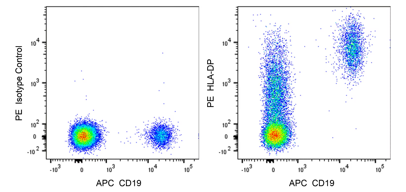
    Two-color flow cytometric analysis of Human HLA-DP expression on human peripheral blood lymphocytes. Human whole blood was stained with APC Mouse Anti-Human CD19 antibody (Cat. No. 555415/561742) and either PE Mouse IgG1, κ Isotype Control (Cat. No. 554680; Left Plot) or PE Mouse Anti-Human HLA-DP antibody (Cat. No. 566825; Right Plot). The erythrocytes were lysed with BD Pharm Lyse™ Lysing Buffer (Cat. No. 555899). A two-color pseudocolor density plot showing the correlated expression of CD19 versus HLA-DP (or Ig Isotype control staining) was derived from gated events with the forward and side-light scatter characteristics of viable lymphocytes. Flow cytometry and data analysis were performed using a BD LSRFortessa™ Cell Analyzer System and FlowJo™ software.

    声明 :本官网所有报价均为常温或者蓝冰运输价格,如有产品需要干冰运输,需另外加收干冰运输费。
    货号:
    566825
    一键复制
    询价
    供应商现货
    100Tst
    选择数量
    当前规格1件起购 
    配送至
    预计5-6个工作日送达,快递: 免运费,若需干冰额外收费