


1/2

品牌: BD Pharmingen
 下载产品说明书 下载SDS
 用小程序,查商品更便捷


 收藏
收藏
 对比
对比 咨询
咨询反应种属:
Human (QC Testing), Rhesus, Cynomolgus, Baboon (Tested in Development), Dog (Reported)
Human (QC Testing), Rhesus, Cynomolgus, Baboon (Tested in Development), Dog (Reported)
来源宿主:
Mouse IgG2a, κ
Mouse IgG2a, κ
产品介绍
产品信息
抗原名称
HLA-DR

宿主
Mouse IgG2a, κ
简单描述
The G46-6 monoclonal antibody specifically binds to HLA-DR, a major histocompatibility complex (MHC) class II antigen. HLA-DR antigens are encoded by genes within the Human Leukocyte Antigen (HLA) Complex located on chromosome 6. HLA-DR is a transmembrane heterodimeric glycoprotein composed of an α chain (36 kDa) and a β subunit (27 kDa) expressed primarily on antigen presenting cells: B cells, dendritic cells, monocytes, macrophages, and thymic epithelial cells. HLA-DR is also expressed on activated T cells. This molecule plays a major role in mediating cellular interactions during antigen presentation to CD4-positive T cells.
商品描述
G46-6
The G46-6 monoclonal antibody specifically binds to HLA-DR, a major histocompatibility complex (MHC) class II antigen. HLA-DR antigens are encoded by genes within the Human Leukocyte Antigen (HLA) Complex located on chromosome 6. HLA-DR is a transmembrane heterodimeric glycoprotein composed of an α chain (36 kDa) and a β subunit (27 kDa) expressed primarily on antigen presenting cells: B cells, dendritic cells, monocytes, macrophages, and thymic epithelial cells. HLA-DR is also expressed on activated T cells. This molecule plays a major role in mediating cellular interactions during antigen presentation to CD4-positive T cells.
同种型
Mouse IgG2a, κ
克隆号
克隆 G46-6 (also known as L243) (RUO)
浓度
1.0 mg/ml
产品详情
NA/LE
NA/LE refers to the culture and purification methods and buffer used to produce purified antibodies with no azide and low endotoxin: Aqueous buffered solution containing no preservative, 0.2µm sterile filtered. Endotoxin level is ≤0.01 EU/µg (≤0.001 ng/µg) of protein as determined by the LAL assay.NA/LE are perfectly suited to be used in culture or in vivo (for nonhuman studies) for functional assays — blocking, neutralizing, activation or depletion — where the presence of azide may damage cells or exogenous endotoxin may signal or activate cells.
应用
实验应用
Flow cytometry (Routinely Tested), Functional assay (Tested During Development)
反应种属
Human (QC Testing), Rhesus, Cynomolgus, Baboon (Tested in Development), Dog (Reported)
目标/特异性
HLA-DR
背景
别名
MHC class II antigen; HLA class II histocompatibility antigen
制备和贮存
存储溶液
No azide/low endotoxin: Aqueous buffered solution containing no preservative, 0.2µm sterile filtered. Endotoxin level is ≤0.01 EU/µg (≤0.001 ng/µg) of protein as determined by the LAL assay.
保存方式
No azide/low endotoxin: Aqueous buffered solution containing no preservative, 0.2µm sterile filtered. Endotoxin level is ≤0.01 EU/µg (≤0.001 ng/µg) of protein as determined by the LAL assay.
文献
文献
研发参考(13)
1. Barclay NA, Brown MH, Birkeland ML, et al, ed. The Leukocyte Antigen FactsBook. San Diego, CA: Academic Press; 1997.
2. Dieckmann D, Plottner H, Berchtold S, Berger T, Schuler G. Ex vivo isolation and characterization of CD4(+)CD25(+) T cells with regulatory properties from human blood. J Exp Med. 2001; 193(11):1303-1310. (Biology).
3. Herodin F, Thullier P, Garin D, Drouet M. Nonhuman primates are relevant models for research in hematology, immunology and virology. Eur Cytokine Netw. 2005; 16(2):104-116. (Biology).
4. Ibisch C, Pradal G, Bach JM, Lieubeau B. Functional canine dendritic cells can be generated in vitro from peripheral blood mononuclear cells and contain a cytoplasmic ultrastructural marker.. J Immunol Methods. 2005; 298(1-2):175-82. (Clone-specific).
5. Kitani A, Chua K, Nakamura K, Strober W. Activated self-MHC-reactive T cells have the cytokine phenotype of Th3/T regulatory cell 1 T cells. J Immunol. 2000; 165(2):691-702. (Clone-specific).
6. Moran TP, Collier M, McKinnon KP, Davis NL, Johnston RE, Serody JS. A novel viral system for generating antigen-specific T cells. J Immunol. 2008; 175(5):3431-3438. (Clone-specific).
7. Pawelec G, Ziegler A, Wernet P. Dissection of human allostimulatory determinants with cloned T cells: stimulation inhibition by monoclonal antibodies TU22, 34, 35, 36, 37, 39, 43, and 58 against distinct human MHC class II molecules. Hum Immunol. 1985; 12(3):165-176. (Biology).
8. Pawelec GP, Shaw S, Ziegler A, Muller C, Wernet P. Differential inhibition of HLA-D- or SB-directed secondary lymphoproliferative responses with monoclonal antibodies detecting human Ia-like determinants. J Immunol. 1982; 129(3):1070-1075. (Biology).
9. Podolin PL, Bolognese BJ, Carpenter DC, et al. Inhibition of invariant chain processing, antigen-induced proliferative responses, and the development of collagen-induced arthritis and experimental autoimmune encephalomyelitis by a small molecule cysteine protease inhibitor. J Immunol. 2008; 180(12):7989-8003. (Biology).
10. Sorg RV, Kogler G, Wernet P. Identification of cord blood dendritic cells as an immature CD11c- population. Blood. 1999; 93(7):2302-2307. (Biology).
11. Ziegler A, Heinig J, Muller C, et al. Analysis by sequential immunoprecipitations of the specificities of the monoclonal antibodies TU22,34,35,36,37,39,43,58 and YD1/63.HLK directed against human HLA class II antigens. Immunobiology. 1986; 171(1-2):77-92. (Biology).
12. Ziegler A, Uchańska-Ziegler B, Zeuthen J, Wernet P. HLA antigen expression at the single cell level on a K562 X B cell hybrid: an analysis with monoclonal antibodies using bacterial binding assays.. Somatic Cell Genet. 1982; 8(6):775-89. (Biology).
13. Zola H. Leukocyte and stromal cell molecules : the CD markers. Hoboken, N.J.: Wiley-Liss; 2007.
参考图片
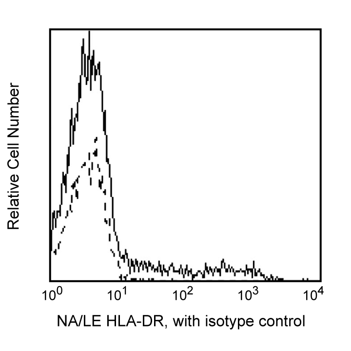
Flow cytometric analysis of HLA-DR on human lysed whole blood. Human whole blood was lysed with BD FACS™ Lysing Solution (Cat. No. 349202) and stained with Purified NA/LE Mouse IgG2a, κ Isotype Control (Cat. No. 554656; dashed line histogram) or with Purified NA/LE Mouse Anti-Human HLA-DR (Cat. No. 555809; solid line histogram). Secondary staining was carried out with FITC Goat Anti-Mouse IgG/IgM (Cat. No. 555988). Fluorescent histograms showing expression of HLA-DR (or Ig isotype staining) were derived from gated events based on forward and side light scattering characteristics for intact lymphocytes. Flow cytometry was performed on a BD FACScan™ system.
声明 :本官网所有报价均为常温或者蓝冰运输价格,如有产品需要干冰运输,需另外加收干冰运输费。

 危险品化学品经营许可证(不带存储) 许可证编号:沪(杨)应急管危经许[2022]202944(QY)
  营业执照(三证合一)