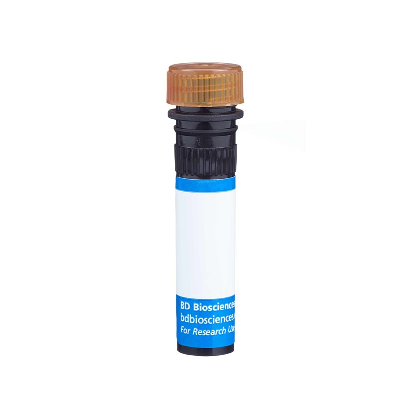


1/2

品牌: BD Pharmingen
 下载产品说明书
 用小程序,查商品更便捷


 收藏
收藏
 对比
对比 咨询
咨询反应种属:
Human (QC Testing), Rhesus, Cynomolgus, Baboon (Tested in Development), Dog (Reactivity Confirmed in Development)
Human (QC Testing), Rhesus, Cynomolgus, Baboon (Tested in Development), Dog (Reactivity Confirmed in Development)
来源宿主:
Mouse IgG2a, κ
Mouse IgG2a, κ
产品介绍
产品信息
耦联标记
PerCP-Cy5.5

抗原名称
HLA-DR

宿主
Mouse IgG2a, κ

简单描述
The G46-6 monoclonal antibody specifically binds to HLA-DR, a major histocompatibility complex (MHC) class II antigen. HLA-DR antigens are encoded by genes within the Human Leukocyte Antigen (HLA) Complex located on chromosome 6. HLA-DR is a transmembrane heterodimeric glycoprotein composed of an α chain (36 kDa) and a β subunit (27 kDa) expressed primarily on antigen presenting cells: B cells, dendritic cells, monocytes, macrophages, and thymic epithelial cells. HLA-DR is also expressed on activated T cells. This molecule plays a major role in mediating cellular interactions during antigen presentation to CD4-positive T cells.

商品描述
The G46-6 monoclonal antibody specifically binds to HLA-DR, a major histocompatibility complex (MHC) class II antigen. HLA-DR antigens are encoded by genes within the Human Leukocyte Antigen (HLA) Complex located on chromosome 6. HLA-DR is a transmembrane heterodimeric glycoprotein composed of an α chain (36 kDa) and a β subunit (27 kDa) expressed primarily on antigen presenting cells: B cells, dendritic cells, monocytes, macrophages, and thymic epithelial cells. HLA-DR is also expressed on activated T cells. This molecule plays a major role in mediating cellular interactions during antigen presentation to CD4-positive T cells.

同种型
Mouse IgG2a, κ

克隆号
G46-6

产品详情
PerCP-Cy5.5
PerCP-Cy5.5 dye is part of the BD blue family of dyes. This tandem fluorochrome is comprised of a fluorescent protein complex (PerCP) with an excitation maximum (Ex Max) of 482 nm and an acceptor dye with an emission maximum (Em Max) at 676 nm. PerCP-Cy5 is designed to be excited by the blue laser (488-nm) and detected using an optical filter centered near 680 nm (e.g., a 695/40 nm bandpass filter). The donor dye can be partially excited by the Violet (405-nm) laser resulting in cross-laser excitation and fluorescence spillover. Please ensure that your instrument’s configurations (lasers and optical filters) are appropriate for this dye.

PerCP-Cy5.5
Blue 488 nm
482 nm
676 nm
应用
实验应用
Flow cytometry (Routinely Tested)

推荐用量
5 µl

反应种属
Human (QC Testing), Rhesus, Cynomolgus, Baboon (Tested in Development), Dog (Reactivity Confirmed in Development)

背景
别名
MHC class II antigen; HLA class II histocompatibility antigen

制备和贮存
存储溶液
Aqueous buffered solution containing BSA and ≤0.09% sodium azide.

保存方式
Aqueous buffered solution containing BSA and ≤0.09% sodium azide.
文献
文献
研发参考(13)
1. Barclay NA, Brown MH, Birkeland ML, et al, ed. The Leukocyte Antigen FactsBook. San Diego, CA: Academic Press; 1997.
2. Dieckmann D, Plottner H, Berchtold S, Berger T, Schuler G. Ex vivo isolation and characterization of CD4(+)CD25(+) T cells with regulatory properties from human blood. J Exp Med. 2001; 193(11):1303-1310. (Biology).
3. Herodin F, Thullier P, Garin D, Drouet M. Nonhuman primates are relevant models for research in hematology, immunology and virology. Eur Cytokine Netw. 2005; 16(2):104-116. (Biology).
4. Ibisch C, Pradal G, Bach JM, Lieubeau B. Functional canine dendritic cells can be generated in vitro from peripheral blood mononuclear cells and contain a cytoplasmic ultrastructural marker.. J Immunol Methods. 2005; 298(1-2):175-82. (Clone-specific).
5. Kitani A, Chua K, Nakamura K, Strober W. Activated self-MHC-reactive T cells have the cytokine phenotype of Th3/T regulatory cell 1 T cells. J Immunol. 2000; 165(2):691-702. (Clone-specific).
6. Moran TP, Collier M, McKinnon KP, Davis NL, Johnston RE, Serody JS. A novel viral system for generating antigen-specific T cells. J Immunol. 2008; 175(5):3431-3438. (Clone-specific).
7. Pawelec G, Ziegler A, Wernet P. Dissection of human allostimulatory determinants with cloned T cells: stimulation inhibition by monoclonal antibodies TU22, 34, 35, 36, 37, 39, 43, and 58 against distinct human MHC class II molecules. Hum Immunol. 1985; 12(3):165-176. (Biology).
8. Pawelec GP, Shaw S, Ziegler A, Muller C, Wernet P. Differential inhibition of HLA-D- or SB-directed secondary lymphoproliferative responses with monoclonal antibodies detecting human Ia-like determinants. J Immunol. 1982; 129(3):1070-1075. (Biology).
9. Podolin PL, Bolognese BJ, Carpenter DC, et al. Inhibition of invariant chain processing, antigen-induced proliferative responses, and the development of collagen-induced arthritis and experimental autoimmune encephalomyelitis by a small molecule cysteine protease inhibitor. J Immunol. 2008; 180(12):7989-8003. (Biology).
10. Sorg RV, Kogler G, Wernet P. Identification of cord blood dendritic cells as an immature CD11c- population. Blood. 1999; 93(7):2302-2307. (Biology).
11. Ziegler A, Heinig J, Muller C, et al. Analysis by sequential immunoprecipitations of the specificities of the monoclonal antibodies TU22,34,35,36,37,39,43,58 and YD1/63.HLK directed against human HLA class II antigens. Immunobiology. 1986; 171(1-2):77-92. (Biology).
12. Ziegler A, Uchańska-Ziegler B, Zeuthen J, Wernet P. HLA antigen expression at the single cell level on a K562 X B cell hybrid: an analysis with monoclonal antibodies using bacterial binding assays.. Somatic Cell Genet. 1982; 8(6):775-89. (Biology).
13. Zola H. Leukocyte and stromal cell molecules : the CD markers. Hoboken, N.J.: Wiley-Liss; 2007.

参考图片
Flow cytometric analysis of HLA-DR on human lysed whole blood. Human whole blood was lysed with BD FACS™ Lysing Solution (Cat. No. 349202) and stained with the PerCP-Cy™5.5 Mouse Anti-Human HLA-DR antibody (Cat. No. 560652/552764; unshaded histogram) or with a PerCP-Cy™5.5 Mouse IgG2a, κ isotype control (Cat. No. 550927; shaded histogram). Fluorescent histograms showing expression of HLA-DR (or Ig isotype staining) were derived from gated events based on forward and side light scattering characteristics for intact lymphocytes. Flow cytometry was performed on a BD™ LSR II flow cytometry system.
声明 :本官网所有报价均为常温或者蓝冰运输价格,如有产品需要干冰运输,需另外加收干冰运输费。


 危险品化学品经营许可证(不带存储) 许可证编号:沪(杨)应急管危经许[2022]202944(QY)
  营业执照(三证合一)