


1/2
BUV395 Mouse Anti-Human HLA-DR(G46-6)


品牌: BD Pharmingen
 下载产品说明书
 用小程序,查商品更便捷


 收藏
收藏
 对比
对比 咨询
咨询反应种属:
Human (QC Testing), Rhesus, Cynomolgus, Baboon (Tested in Development), Dog (Reported)
Human (QC Testing), Rhesus, Cynomolgus, Baboon (Tested in Development), Dog (Reported)
来源宿主:
Mouse IgG2a, κ
Mouse IgG2a, κ
产品介绍
产品信息
耦联标记
BUV395

抗原名称
HLA-DR
宿主
Mouse IgG2a, κ
简单描述
The G46-6 monoclonal antibody specifically binds to HLA-DR, a major histocompatibility complex (MHC) class II antigen. HLA-DR antigens are encoded by genes within the Human Leukocyte Antigen (HLA) Complex located on chromosome 6. HLA-DR is a transmembrane heterodimeric glycoprotein composed of an α chain (36 kDa) and a β subunit (27 kDa) expressed primarily on antigen presenting cells: B cells, dendritic cells, monocytes, macrophages, and thymic epithelial cells. HLA-DR is also expressed on activated T cells. This molecule plays a major role in mediating cellular interactions during antigen presentation to CD4-positive T cells.
The antibody was conjugated to BD Horizon™ BUV395 which has been exclusively developed by BD Biosciences as an optimal dye for use on a 355 nm laser equipped instrument. With an Ex Max at 348 nm and an Em Max at 395 nm, this dye has virtually no spillover into any other detector. BD Horizon™ BUV395 can be excited with a 355 nm laser and detected with a 379/28 filter.
商品描述
The G46-6 monoclonal antibody specifically binds to HLA-DR, a major histocompatibility complex (MHC) class II antigen. HLA-DR antigens are encoded by genes within the Human Leukocyte Antigen (HLA) Complex located on chromosome 6. HLA-DR is a transmembrane heterodimeric glycoprotein composed of an α chain (36 kDa) and a β subunit (27 kDa) expressed primarily on antigen presenting cells: B cells, dendritic cells, monocytes, macrophages, and thymic epithelial cells. HLA-DR is also expressed on activated T cells. This molecule plays a major role in mediating cellular interactions during antigen presentation to CD4-positive T cells.
The antibody was conjugated to BD Horizon™ BUV395 which has been exclusively developed by BD Biosciences as an optimal dye for use on a 355 nm laser equipped instrument. With an Ex Max at 348 nm and an Em Max at 395 nm, this dye has virtually no spillover into any other detector. BD Horizon™ BUV395 can be excited with a 355 nm laser and detected with a 379/28 filter.
同种型
Mouse IgG2a, κ
克隆号
G46-6
产品详情
BUV395
The BD Horizon Brilliant™ Ultraviolet 395 (BUV395) Dye is part of the BD Horizon Brilliant™ Ultraviolet family of dyes. This base dye is a polymer fluorochrome with an excitation maximum (Ex Max) of 348-nm and an emission maximum (Em Max) at 395-nm. Driven by BD innovation, BUV395 is designed to be excited by the ultraviolet laser (355-nm) and detected using an optical filter centered near 380-nm (e.g., 379/28-nm bandpass filter). BUV395 is the ideal dye when using only one detector on the ultraviolet laser as it spills into no other detectors and no other fluors spill into it. Please ensure that your instrument’s configurations (lasers and optical filters) are appropriate for this dye.

BUV395
Ultraviolet 355 nm
348 nm
395 nm
应用
实验应用
Flow cytometry (Routinely Tested)
推荐用量
5 µl
反应种属
Human (QC Testing), Rhesus, Cynomolgus, Baboon (Tested in Development), Dog (Reported)
背景
别名
MHC class II antigen; HLA class II histocompatibility antigen
制备和贮存
存储溶液
Aqueous buffered solution containing BSA and ≤0.09% sodium azide.
保存方式
Aqueous buffered solution containing BSA and ≤0.09% sodium azide.
文献
文献
研发参考(5)
1. Dieckmann D, Plottner H, Berchtold S, Berger T, Schuler G. Ex vivo isolation and characterization of CD4(+)CD25(+) T cells with regulatory properties from human blood. J Exp Med. 2001; 193(11):1303-1310. (Clone-specific: Flow cytometry).
2. Ibisch C, Pradal G, Bach JM, Lieubeau B. Functional canine dendritic cells can be generated in vitro from peripheral blood mononuclear cells and contain a cytoplasmic ultrastructural marker.. J Immunol Methods. 2005; 298(1-2):175-82. (Clone-specific).
3. Kitani A, Chua K, Nakamura K, Strober W. Activated self-MHC-reactive T cells have the cytokine phenotype of Th3/T regulatory cell 1 T cells. J Immunol. 2000; 165(2):691-702. (Clone-specific: Flow cytometry).
4. Moran TP, Collier M, McKinnon KP, Davis NL, Johnston RE, Serody JS. A novel viral system for generating antigen-specific T cells. J Immunol. 2008; 175(5):3431-3438. (Clone-specific: Flow cytometry).
5. Sorg RV, Kogler G, Wernet P. Identification of cord blood dendritic cells as an immature CD11c- population. Blood. 1999; 93(7):2302-2307. (Clone-specific: Flow cytometry).
参考图片
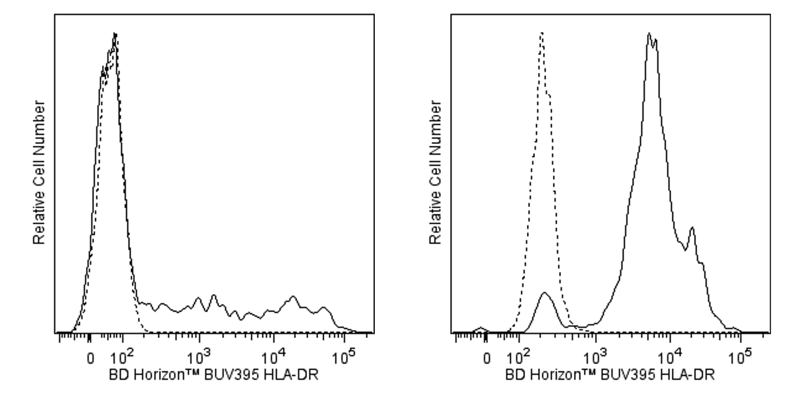
Flow cytometric analysis of HLA-DR expression on human peripheral blood lymphocytes and monocytes. Human whole blood was stained with either BD Horizon™ BUV395 Mouse IgG2a, κ Isotype Control (Cat. No. 563809; dashed line histogram) or BD Horizon™ BUV395 Mouse Anti-Human HLA-DR antibody (Cat. No. 564040/565972; solid line histogram). The erythrocytes were lysed with BD FACS™ Lysing Solution (Cat. No. 349202). The fluorescence histograms were derived from gated events with the forward and side light-scatter characteristics of intact lymphocytes (Left Panel) or monocytes (Right Panel). Flow cytometric analysis was performed using a BD™ LSR II Flow Cytometer System.
声明 :本官网所有报价均为常温或者蓝冰运输价格,如有产品需要干冰运输,需另外加收干冰运输费。

 危险品化学品经营许可证(不带存储) 许可证编号:沪(杨)应急管危经许[2022]202944(QY)
  营业执照(三证合一)