


 下载产品说明书
 下载SDS
下载SDS 用小程序,查商品更便捷


 收藏
收藏
 对比
对比 咨询
咨询Immunohistochemistry(8-25 µg/mL)
Immunocytochemistry(8-25 µg/mL)
Immunohistochemistry(8-25 µg/mL)
Immunocytochemistry(8-25 µg/mL)


Ser2-Leu414
Accession # O75874


Scientific Data
 View Larger
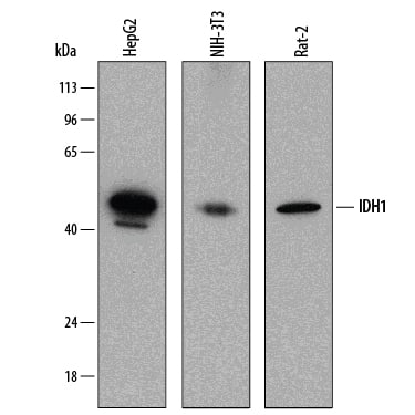
Detection of Human, Mouse, and Rat Isocitrate Dehydrogenase 1/IDH1 by Western Blot. Western blot shows lysates of HepG2 human hepatocellular carcinoma cell line, NIH-3T3 mouse embryonic fibroblast cell line, and Rat-2 rat embryonic fibroblast cell line. PVDF membrane was probed with 0.25 µg/mL of Mouse Anti-Human Isocitrate Dehydrogenase 1/IDH1 Monoclonal Antibody (Catalog # MAB7049) followed by HRP-conjugated Anti-Mouse IgG Secondary Antibody (HAF018). A specific band was detected for Isocitrate Dehydrogenase 1/IDH1 at approximately 46 kDa (as indicated). This experiment was conducted under reducing conditions and using Immunoblot Buffer Group 1.
 View Larger
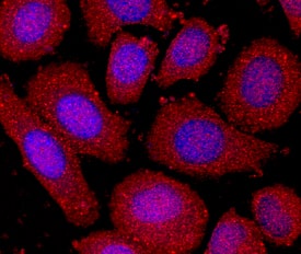
Isocitrate Dehydrogenase 1/IDH1 in SK‑BR‑3 Human Cell Line. Isocitrate Dehydrogenase 1/IDH1 was detected in immersion fixed SK-BR-3 human breast cancer cell line using Mouse Anti-Human Isocitrate Dehydrogenase 1/IDH1 Monoclonal Antibody (Catalog # MAB7049) at 10 µg/mL for 3 hours at room temperature. Cells were stained using the NorthernLights™ 557-conjugated Anti-Mouse IgG Secondary Antibody (red; NL007) and counterstained with DAPI (blue). Specific staining was localized to cytoplasm. View our protocol for Fluorescent ICC Staining of Cells on Coverslips.
 View Larger
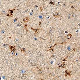
Isocitrate Dehydrogenase 1/IDH1 in Human Brain. Isocitrate Dehydrogenase 1/IDH1 was detected in immersion fixed paraffin-embedded sections of human brain (cortex) using Mouse Anti-Human Isocitrate Dehydrogenase 1/IDH1 Monoclonal Antibody (Catalog # MAB7049) at 15 µg/mL overnight at 4 °C. Before incubation with the primary antibody, tissue was subjected to heat-induced epitope retrieval using Antigen Retrieval Reagent-Basic (CTS013). Tissue was stained using the Anti-Mouse HRP-DAB Cell & Tissue Staining Kit (brown; CTS002) and counterstained with hematoxylin (blue). Specific staining was localized to astrocytes. View our protocol for Chromogenic IHC Staining of Paraffin-embedded Tissue Sections.
 View Larger
Isocitrate Dehydrogenase 1/IDH1 in Rat Brain. Isocitrate Dehydrogenase 1/IDH1 was detected in perfusion fixed frozen sections of rat brain using Mouse Anti-Human Isocitrate Dehydrogenase 1/IDH1 Monoclonal Antibody (Catalog # MAB7049) at 15 µg/mL overnight at 4 °C. Tissue was stained using the NorthernLights™ 557-conjugated Anti-Mouse IgG Secondary Antibody (red; NL007) and counterstained with DAPI (blue). Specific staining was localized to glial cell cytoplasm. View our protocol for Fluorescent IHC Staining of Frozen Tissue Sections.
 View Larger
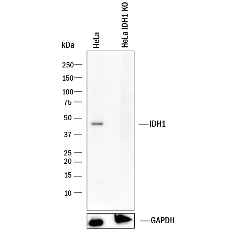
Western Blot Shows Human Isocitrate Dehydrogenase 1/IDH1 Specificity by Using Knockout Cell Line. Western blot shows lysates of HeLa human cervical epithelial carcinoma parental cell line and Isocitrate Dehydrogenase 1/IDH1 knockout HeLa cell line (KO). PVDF membrane was probed with 0.25 µg/mL of Mouse Anti-Human Isocitrate Dehydrogenase 1/IDH1 Monoclonal Antibody (Catalog # MAB7049) followed by HRP-conjugated Anti-Mouse IgG Secondary Antibody (HAF018). A specific band was detected for Isocitrate Dehydrogenase 1/IDH1 at approximately 46 kDa (as indicated) in the parental HeLa cell line, but is not detectable in knockout HeLa cell line. GAPDH (MAB5718) is shown as a loading control. This experiment was conducted under reducing conditions and using Immunoblot Buffer Group 1.
 View Larger
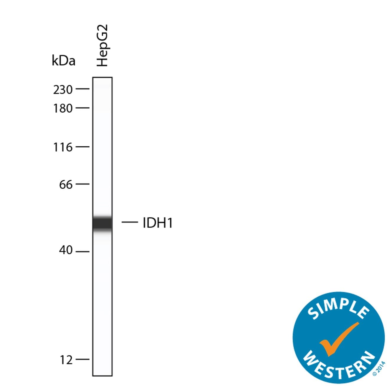
Detection of Human Isocitrate Dehydrogenase 1/IDH1 by Simple WesternTM. Simple Western lane view shows lysates of HepG2 human hepatocellular carcinoma cell line, loaded at 0.2 mg/mL. A specific band was detected for Isocitrate Dehydrogenase 1/IDH1 at approximately 51 kDa (as indicated) using 10 µg/mL of Mouse Anti-Human Isocitrate Dehydrogenase 1/IDH1 Monoclonal Antibody (Catalog # MAB7049). This experiment was conducted under reducing conditions and using the 12-230 kDa separation system.
Human Isocitrate Dehydrogenase 1/IDH1 Antibody Summary
Ser2-Leu414
Accession # O75874
Applications
Please Note: Optimal dilutions should be determined by each laboratory for each application. General Protocols are available in the Technical Information section on our website.
Immunohistochemistry(8-25 µg/mL)
Immunocytochemistry(8-25 µg/mL)


Background: Isocitrate Dehydrogenase 1/IDH1
Isocitrate Dehydrogenase 1 (IDH1) catalyzes the oxidative decarboxylation of isocitrate to alpha ‑ketoglutarate. There are two subclasses in the IDH family, one of them utilizing NADP+ as the electron acceptor and the other using NAD+ (1). The protein encoded by this gene is the NADP+-dependent isocitrate dehydrogenase found in the cytoplasm and peroxisomes. In peroxisomes, IDH1 generates the NADPH required for intraperoxisomal reduction reactions. Mutations of Arg132 of human IDH1 result in a reduced ability of the enzyme to convert isocitrate to alpha ‑ketoglutarate, but the enzyme acquires the ability to generate 2-hydroxyglutarate (2HG) from alpha ‑ketoglutarate (2). Elevated levels of the metabolite 2HG are associated with a high risk of malignant brain tumors. Arg132 mutations of IDH1 are common in high‑grade gliomas, but not in other types of tumors (3).
- Nekrutenko, A. et al. (1998) Mol. Biol. Evol. 15:1674.
- Dang, L. et al. (2009) Nature 462:739.
- Bleeker, F.E. et al. (2009) Hum. Mutat. 30:7.


Preparation and Storage
- 12 months from date of receipt, -20 to -70 °C as supplied.
- 1 month, 2 to 8 °C under sterile conditions after reconstitution.
- 6 months, -20 to -70 °C under sterile conditions after reconstitution.
参考图片
Detection of Human, Mouse, and Rat Isocitrate Dehydrogenase 1/IDH1 by Western Blot. Western blot shows lysates of HepG2 human hepatocellular carcinoma cell line, NIH‑3T3 mouse embryonic fibroblast cell line, and Rat‑2 rat embryonic fibroblast cell line. PVDF membrane was probed with 0.25 µg/mL of Mouse Anti-Human Isocitrate Dehydrogenase 1/IDH1 Monoclonal Antibody (Catalog # MAB7049) followed by HRP-conjugated Anti-Mouse IgG Secondary Antibody (Catalog # HAF018). A specific band was detected for Isocitrate Dehydrogenase 1/IDH1 at approximately 46 kDa (as indicated). This experiment was conducted under reducing conditions and using Immunoblot Buffer Group 1.
Isocitrate Dehydrogenase 1/IDH1 in SK‑BR‑3 Human Cell Line. Isocitrate Dehydrogenase 1/IDH1 was detected in immersion fixed
SK‑BR‑3 human breast cancer cell line using Mouse Anti-Human Isocitrate Dehydrogenase 1/IDH1 Monoclonal Antibody (Catalog # MAB7049) at 10 µg/mL for 3 hours at room temperature. Cells were stained using the NorthernLights™ 557-conjugated Anti-Mouse IgG Secondary Antibody (red; Catalog # NL007) and counterstained with DAPI (blue). Specific staining was localized to cytoplasm. View our protocol for Fluorescent ICC Staining of Cells on Coverslips.
Isocitrate Dehydrogenase 1/IDH1 in Human Brain. Isocitrate Dehydrogenase 1/IDH1 was detected in immersion fixed paraffin-embedded sections of human brain (cortex) using Mouse Anti-Human Isocitrate Dehydrogenase 1/IDH1 Monoclonal Antibody (Catalog # MAB7049) at 15 µg/mL overnight at 4 °C. Before incubation with the primary antibody, tissue was subjected to heat-induced epitope retrieval using Antigen Retrieval Reagent-Basic (Catalog # CTS013). Tissue was stained using the Anti-Mouse HRP-DAB Cell & Tissue Staining Kit (brown; Catalog # CTS002) and counterstained with hematoxylin (blue). Specific staining was localized to astrocytes. View our protocol for Chromogenic IHC Staining of Paraffin-embedded Tissue Sections.
Isocitrate Dehydrogenase 1/IDH1 in Rat Brain. Isocitrate Dehydrogenase 1/IDH1 was detected in perfusion fixed frozen sections of rat brain using Mouse Anti-Human Isocitrate Dehydrogenase 1/IDH1 Monoclonal Antibody (Catalog # MAB7049) at 15 µg/mL overnight at 4 °C. Tissue was stained using the NorthernLights™ 557-conjugated Anti-Mouse IgG Secondary Antibody (red; Catalog # NL007) and counterstained with DAPI (blue). Specific staining was localized to glial cell cytoplasm. View our protocol for Fluorescent IHC Staining of Frozen Tissue Sections.



 危险品化学品经营许可证(不带存储) 许可证编号:沪(杨)应急管危经许[2022]202944(QY)
  营业执照(三证合一)