
 下载产品说明书
 下载SDS
下载SDS 用小程序,查商品更便捷


 收藏
收藏
 对比
对比 咨询
咨询


Scientific Data
 View Larger
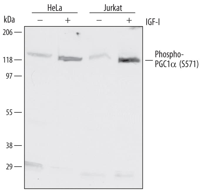
Detection of Human Phospho-PGC1 alpha (S571) by Western Blot. Western blot shows lysates of HeLa human cervical epithelial carcinoma cell line and Jurkat human acute T cell leukemia cell line untreated (-) or treated (+) with 10 nm Recombinant Human IGF-I (Catalog # 291-G1) for 1 hour. PVDF Membrane was probed with 1 µg/mL of Rabbit Anti-Human Phospho-PGC1a (S571) Antigen Affinity-purified Polyclonal Antibody (Catalog # AF6650) followed by HRP-conjugated Anti-Rabbit IgG Secondary Antibody (Catalog # HAF008). A specific band was detected for Phospho-PGC1a (S571) at approximately 120 kDa (as indicated). This experiment was conducted under reducing conditions and using Immunoblot Buffer Group 1.
Human Phospho-PGC1 alpha (S571) Antibody Summary
Applications
Please Note: Optimal dilutions should be determined by each laboratory for each application. General Protocols are available in the Technical Information section on our website.


Background: PGC1 alpha
PGC-1 alpha (PPAR-gamma coactivator 1; also LEM6) is a 97-120 kDa member of the PGC-1 family of proteins. It is expressed in select cell types, including brown adipocytes, skeletal muscle and hepatocytes. PGC-1 alpha participates in both RNA processing and transcriptional coactivation in conjunction with multiple nuclear hormone receptors such as PPAR gamma, RAR and TR. Human PCG-1 alpha is 798 amino acids (aa) in length. It contains an LxxLL nuclear receptor binding motif (aa 144-148), one PPAR-gamma interaction domain (aa 293-339), two NLSs and an RNA binding/processing region (aa 566-710). PGC-1 alpha activity is regulated by phosphorylation. AMPK is known to phosphorylate Thr178 and Ser539, promoting cotranscriptional activity. Conversely, Akt-mediated phosphorylation at Ser571 is reported to downregulate PGC-1 alpha activity. This latter effect is achieved by an initial Ser571 phosphorylation, followed by GCN5 binding and subsequent PCG-1 alpha acetylation that promotes PGC-1 alpha dissociation from target gene promoters.


Preparation and Storage
- 12 months from date of receipt, -20 to -70 °C as supplied.
- 1 month, 2 to 8 °C under sterile conditions after reconstitution.
- 6 months, -20 to -70 °C under sterile conditions after reconstitution.
参考图片
Detection of Human Phospho-PGC1 alpha (S571) by Western Blot. Western blot shows lysates of HeLa human cervical epithelial carcinoma cell line and Jurkat human acute T cell leukemia cell line untreated (-) or treated (+) with 10 nm Recombinant Human IGF‑I (Catalog # 291-G1) for 1 hour. PVDF Membrane was probed with 1 µg/mL of Rabbit Anti-Human Phospho-PGC1 alpha (S571) Antigen Affinity-purified Polyclonal Antibody (Catalog # AF6650) followed by HRP-conjugated Anti-Rabbit IgG Secondary Antibody (Catalog # HAF008). A specific band was detected for Phospho-PGC1 alpha (S571) at approximately 120 kDa (as indicated). This experiment was conducted under reducing conditions and using Immunoblot Buffer Group 1.


 危险品化学品经营许可证(不带存储) 许可证编号:沪(杨)应急管危经许[2022]202944(QY)
  营业执照(三证合一)