

 下载产品说明书
 用小程序,查商品更便捷


 收藏
收藏
 对比
对比 咨询
咨询49-72kDa (Reducing)
49-72kDa (Reducing)
>95% by SDS-PAGE
>95% by SDS-PAGE

Gln28-Glu480, with C-terminal Human IgG1 Fc
QTSVSPSKVILPRGGSVLVTCSTSCDQPKLLGIETPLPKKELLLPGNNRKVYELSNVQEDSQPMCYSNCPDGQSTAKTFLTVYWTPERVELAPLPSWQPVGKNLTLRCQVEGGAPRANLTVVLLRGEKELKREPAVGEPAEVTTTVLVRRDHHGANFSCRTELDLRPQGLELFENTSAPYQLQTFVLPATPPQLVSPRVLEVDTQGTVVCSLDGLFPVSEAQVHLALGDQRLNPTVTYGNDSFSAKASVSVTAEDEGTQRLTCAVILGNQSQETLQTVTIYSFPAPNVILTKPEVSEGTEVTVKCEAHPRAKVTLNGVPAQPLGPRAQLLLKATPEDNGRSFSCSATLEVAGQLIHKNQTRELRVLYGPRLDERDCPGNWTWPENSQQTPMCQAWGNPLPELKCLKDGTFPLPIGESVTVTRDLEGTYLCRARSTQGEVTRKVTVNVLSPRYEIEGRMDPKSSDKTHTCPPCPAPELLGGPSVFLFPPKPKDTLMISRTPEVTCVVVDVSHEDPEVKFNWYVDGVEVHNAKTKPREEQYNSTYRVVSVLTVLHQDWLNGKEYKCKVSNKALPAPIEKTISKAKGQPREPQVYTLPPSRDELTKNQVSLTCLVKGFYPSDIAVEWESNGQPENNYKTTPPVLDSDGSFFLYSKLTVDKSRWQQGNVFSCSVMHEALHNHYTQKSLSLSPGK

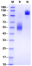
49-72kDa (Reducing)

>95% by SDS-PAGE


ICAM-1 is a cell-surface glycoprotein and a member of the immunoglobulin (Ig) protein superfamily. ICAM-1 mediates the interaction between keratinocytes and leukocytes. ICAM-1 facilitates a variety of both afferent and efferent immune responses that require intercellular contact and collaboration, including T helper responses, T-dependent B cell responses, antigen-induced T cell proliferation, cell-mediated cytotoxicity, and the binding of leukocytes to vascular endothelium prior to extravasation into inflamed tissue. Pro-inflammatory cytokines interferon-γ and TNF-α stimulate the expression of keratinocyte ICAM-1 and this up-regulation is blocked both in vivo and in vitro if keratinocytes are irradiated with UVB prior to cytokine treatment. In contrast, ICAM-2 is expressed primarily on endothelium and leukocytes (except neutrophils), and its expression generally is not responsive to cytokines. ICAM-3 predominates on thymocytes and resting lymphocytes, which are low in ICAM-1. When neutrophils are activated, they shed the majority of their ICAM-3 into the medium by proteolytic cleavage. In some reports, the expression of ICAM-1 has been positively correlated with a more aggressive tumor phenotype and metastatic potential. For instance, the invasiveness of breast cancer cells has been positively correlated with the expression of ICAM-1. Also, it has been suggested that an ICAM-1–ICAM-1 homophilic interaction between breast cancer cells and mesenchymal stem cells in bone marrow mediates the metastatic expansion of cancer cells, displacing hematopoietic stem cells from their niche.

Reconstitute at 0.1-1 mg/ml according to the size in ultrapure water after rapid centrifugation.

- 12 months from date of receipt, -20 to -70 °C as supplied.
- 6 months, -20 to -70 °C under sterile conditions after reconstitution.
- 1 week, 2 to 8 °C under sterile conditions after reconstitution.
- Please avoid repeated freeze-thaw cycles.
参考图片
2μg (R: reducing conditions, N: non-reducing conditions).
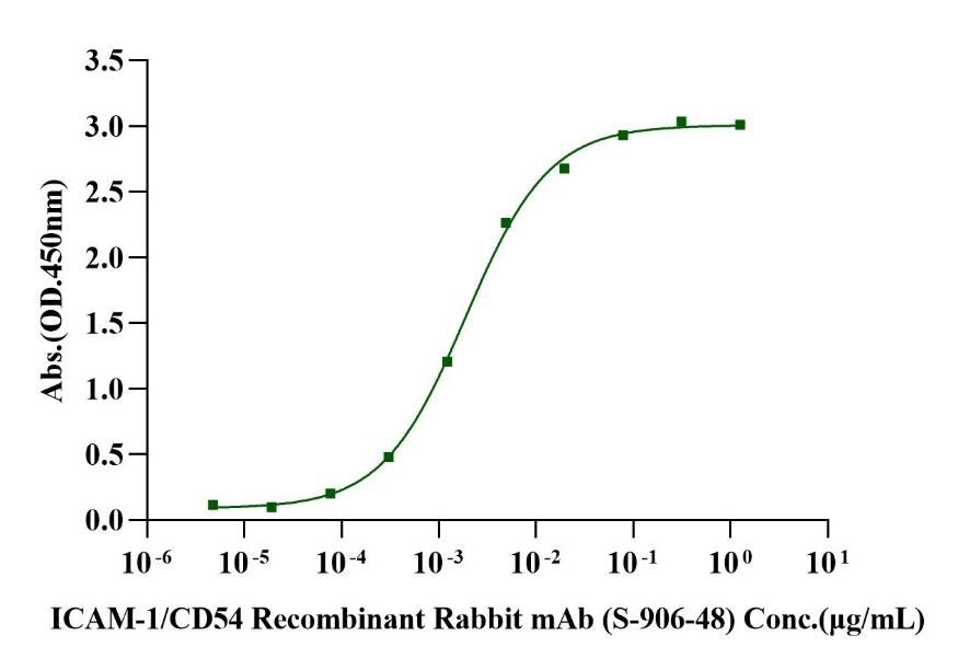
Immobilized ICAM-1/CD54 Fc Chimera, Human (Cat. No. UA010156) at 2.0μg/mL (100μL/well) can bind ICAM-1/CD54 Recombinant Rabbit mAb (S-906-48) (S-1233-25) (Cat. No. S0B0795) with EC50 of 1.62-2.20ng/mL.


 危险品化学品经营许可证(不带存储) 许可证编号:沪(杨)应急管危经许[2022]202944(QY)
  营业执照(三证合一)