


 用小程序,查商品更便捷


Product Usage Information
The optimal dilution of the anti-species antibody should be determined for each primary antibody by titration. However, a final dilution of 1:500 – 1:2000 should yield acceptable results for immunofluorescent and flow cytometry assays.
Specificity/Sensitivity
Species Reactivity:
Mouse
参考图片
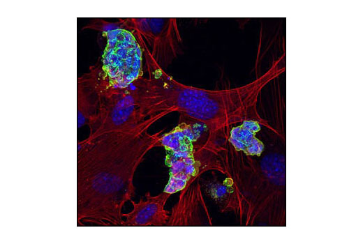
Confocal immunofluorescent analysis of mouse embryonic stem cells growing on mouse embryonic fibroblast (MEF) feeder cells using SSEA1 (MC480) Mouse mAb #4744 detected with anti-Mouse IgG (H+L), F(ab')2 Fragment (Alexa Fluor® 488 Conjugate) (green). Actin filaments have been labeled with DY-554 phalloidin (red). Blue pseudocolor = DRAQ5® #4084 (fluorescent DNA dye).激光共聚焦免疫荧光分析生长在小鼠胚胎成纤维饲养层细胞上的小鼠胚胎干细胞,所用抗体为SSEA1 (MC480) Mouse mAb #4744 检测抗体为anti-Mouse IgG (H+L), F(ab’)2 Fragment (Alexa Fluor ® 488 Conjugate) (绿色)。 肌动蛋白丝采用DY-554 phalloidin (红色)标记。蓝色伪彩为荧光DNA染料,信息为DRAQ5 ® #4084。
Flow cytometric analysis of untreated Jurkat cells using Akt (5G3) Mouse mAb #2966 detected with Anti-Mouse IgG (H+L), F(ab')2 Fragment (Alexa Fluor® 488 Conjugate) (green) compared to a nonspecific negative control antibody (red).流式细胞术分析Jurkat细胞,所用抗体为Akt (5G3) Mouse mAb #2966,检测抗体为Anti-Mouse IgG (H+L), F(ab’)2Fragment (Alexa Fluor ® 488 Conjugate) (绿色) ,与非特异性阴性对照抗体(红色)作对比。
High content analysis of A439 cells exposed to varying concentrations of caffeine for 30 min prior to and 1.5 hr following a 100 mJ UV-treatment. With increasing concentrations of caffeine, a significant decrease (~2.5 fold) in phospho-p53 signal as compared to the UV-treated control was observed. When using phospho-p53 as a measurement, the IC50 of this compound was 2.95 mM. Data was generated on the Acumen® HCS platform using Anti-Mouse IgG (H+L), F(ab')2 Fragment (Alexa Fluor® 488 Conjugate).高通量分析A439细胞,该细胞暴露于咖啡因30分钟,然后用100J UV处理1.5个小时。与UV处理的对照组相比较,随着咖啡因浓度的增加, phospho-p53信号显著降低(约2.5倍)。当采用 phospho-p53检测时,IC50为2.95mM。数据时以Acumen ® HCS为平台生成的,所用抗体为Anti-Mouse IgG (H+L), F(ab’)2 Fragment (Alexa Fluor ® 488 Conjugate)。

 危险品化学品经营许可证(不带存储) 许可证编号:沪(杨)应急管危经许[2022]202944(QY)
  营业执照(三证合一)