


 用小程序,查商品更便捷


Product Usage Information
The optimal dilution of the anti-species antibody should be determined for each primary antibody by titration. However, a final dilution of 1:500 - 1:2000 should yield acceptable results for immunofluorescent assays.
Specificity/Sensitivity
Species Reactivity:
Mouse
参考图片
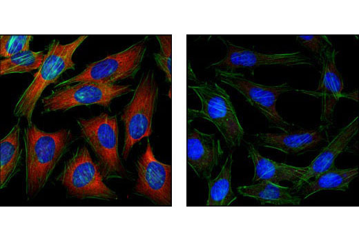
Confocal immunofluorescent analysis of HeLa cells using S6 Ribosomal Protein (54D2) Mouse mAb #2317 detected with Anti-Mouse IgG (H+L), F(ab')2 Fragment (Alexa Fluor® 555 Conjugate) (red, left) compared to an isotype control (right). Actin filaments have been labeled with fluorescein phalloidin. Blue pseudocolor = DRAQ5® #4084 (fluorescent DNA dye).激光共聚焦免疫荧光分析HeLa细胞,所用抗体为S6 Ribosomal Protein (54D2) Mouse mAb #2317,检测抗体为anti-Mouse IgG (H+L), F(ab’)2 Fragment (Alexa Fluor ® 555 Conjugate) (上图),与同型对照(下图)作比较。 肌动蛋白丝采用fluorescein phalloidi标记。蓝色伪彩为荧光DNA染料,信息为DRAQ5 ® #4084。
High content analysis of C2C12 cells exposed to varying concentrations of LY294002 #9901 for 2 hours. With increasing concentrations of LY294002, a significant decrease (~20 fold) in phospho-S6 Ribosomal protein (Ser235/236) signal as compared to the untreated control was observed. When using phospho-S6 Ribosomal protein as a measurement, the IC50 of this compound was 2.3 μM. Data was generated on the Acumen® HCS platform using Anti-Mouse IgG (H+L), F(ab')2 Fragment (Alexa Fluor® 555 Conjugate).高通量分析C2C12细胞,该细胞暴露于LY294002 #9901 2个小时。与未处理的对照组相比较,随着LY294002 浓度的增加phospho-S6 Ribosomal protein (Ser235/236)信号显著降低(约20倍)。当采用 phospho-S6 Ribosomal protein 检测时,IC50为2.3mM。数据时以Acumen ® HCS为平台生成的,所用抗体为Anti-Mouse IgG (H+L), F(ab’)2 Fragment (Alexa Fluor ® 555 Conjugate)。

 危险品化学品经营许可证(不带存储) 许可证编号:沪(杨)应急管危经许[2022]202944(QY)
  营业执照(三证合一)