


 用小程序,查商品更便捷


Product Usage Information
The optimal dilution of the anti-species antibody should be determined by the user. However, the final dilutions below should yield acceptable results for the respective applications.Fluorescent western blotting: 1:30000In-Cell Western: 1:1000
Specificity/Sensitivity
Species Reactivity:
Rabbit
参考图片
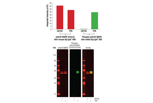
Western blot analysis of Jurkat cell lysates (#9194) treated with either U0126 (MEK 1/2 inhibitor) #9903 or TPA (12-O-Tetradecanoylphorbol-13-Acetate) #4174, using Phospho-p44/42 MAPK (Erk1/2) (Thr202/204) (D13.14.4E) XP® Rabbit mAb #4370 detected with Anti-rabbit IgG (H+L) (DyLight® 800 Conjugate) (green) and p44/42 MAPK (Erk1/2) (3A7) Mouse mAb #9107 detected with Anti-mouse IgG (H+L) (DyLight® 680 Conjugate) #5470 (red). The array image pixel intensities obtained using a LI-COR® Biosciences Odyssey® Infrared Imaging System are shown in the upper panel while corresponding fluorescent western blots are shown in the lower panel.Western blot分析U0126 (MEK 1/2 inhibitor) #9903 或 TPA (12-十四酸佛波酯-13-乙酸盐) #4174处理的Jurkat细胞(#9194)提取物。所用抗体有Phospho-p44/42 MAPK (Erk1/2) (Thr202/204) (D13.14.4E) XP™ Rabbit mAb #4370 ,检测用Anti-rabbit IgG (H+L (DyLight ® 800 Conjugate) #5151(绿色)和p44/42 MAPK (Erk1/2) (3A7) Mouse mAb #9107,检测用Anti-mouse IgG (H+L) (DyLight ® 680 Conjugate) (红色)。上图一系列的像素强度是用LI-COR ® Biosciences Odyssey ® Infrared 成像系统中获得的;下图为相对应的荧光western blots结果。
In-Cell Western™ analysis of A549 cells exposed to varying concentrations of U0126 (MEK1/2 Inhibitor) #9903 for 3 hours, followed by TPA (Phorbol-12-Myristate-13-Acetate) #9905 stimulation for 30 minutes. With increasing concentrations of U0126, a significant decrease (~5 fold) in Phospho p44/42 MAPK (Erk1/2) (Thr202/Tyr204) (D13.14.4E) XP® Rabbit mAb #4370 signal as compared to the TPA-stimulated control was observed. When using phospho-Erk as a measurement, the IC50 of this compound was 2.8 μM. Data and images were generated on the LI-COR® Biosciences Odyssey® Infrared Imaging System using Anti-rabbit IgG (H+L) (DyLight® 800 Conjugate). DRAQ5® #4084 (fluorescent DNA dye - red) is used for normalization.In-Cell Western™分析暴露于不同浓度的U0126 (MEK1/2 抑制剂) #9903的A549细胞,暴露时间为3小时,然后用TPA(12-十四酸佛波酯-13-乙酸盐)刺激30分钟。 与TPA刺激的对照组相比较,随着U0126刺激浓度的增加, Phospho-p44/42 MAPK (Erk1/2) (Thr202/Tyr204 (D13.14.4E) XP™ Rabbit mAb #4370的信号与TPA刺激的对照组相比较显著降低(约5倍)。当用phospho-Erk检测时,复合物的IC50为2.8uM。数据和照片是在LI-COR ® Biosciences Odyssey ® Infrared 成像系统中生成的,所用抗体为Anti-rabbit IgG (H+L) (DyLight ® 800 Conjugate)。 所用标准化抗体是DRAQ5 ® #4084 (荧光DNA 红色染料) 。

 危险品化学品经营许可证(不带存储) 许可证编号:沪(杨)应急管危经许[2022]202944(QY)
  营业执照(三证合一)