Anti-Mouse IgG1 Magnetic Particles - DM(A85-1)
1/3

Anti-Mouse IgG1 Magnetic Particles - DM(A85-1)

分享
品牌: BD Pharmingen
pdf 下载产品说明书
用小程序,查商品更便捷
收藏
对比对比
咨询咨询
    反应种属:
    Mouse (QC Testing)
    Mouse (QC Testing)
    来源宿主:
    Rat LOU, also known as Louvain, LOU/C, LOU/M IgG1, κ
    Rat LOU, also known as Louvain, LOU/C, LOU/M IgG1, κ
    展开
    产品介绍
    产品信息
    耦联标记
    Magnetic Bead
    宿主
    Rat LOU, also known as Louvain, LOU/C, LOU/M IgG1, κ
    免疫原
    Pooled Mouse IgG1
    简单描述
    BD IMag™ Anti-Mouse IgG1 Magnetic Particles -DM are magnetic nanoparticles conjugated with a monoclonal antibody. The A85-1 antibody clone has been reported to react specifically with mouse IgG1 of Igh-Ca and Igh-Cb haplotypes, but not other Ig isotypes. These particles are optimized for the positive selection or depletion of leukocyte subpopulations labelled with mouse IgG1 antibodies using the BD IMag™ Cell Separation Magnet. Leukocytes are labelled with BD IMag™ Anti-Mouse IgG1 Magnetic Particles - DM according to the Magnetic Labeling and Separation Protocol.  In brief, cells are labeled with a mouse IgG1 antibody that recognizes the subpopulation of interest. After washing away excess antibody, BD IMag™ Anti-Mouse IgG1 Magnetic Particles -DM are added to the cell suspension and bind the mouse IgG1 antibody on the cells. This labeled cell suspension is then placed within the magnetic field of the BD IMag™ Cell Separation Magnet (Cat.No. 552311). Positive selection or depletion is then performed. Labeled cells migrate toward the magnet (positive fraction), leaving the unlabeled cells in suspension so they can be drawn off (depleted or negative fraction). The tube is then removed from the magnetic field for resuspension of the positive fraction. The magnetic separation steps are diagrammed in the Depletion and Positive Selection Flow Charts. After the positive fraction has been washed, the small size of the magnetic particles allows the positive fraction to be further evaluated in downstream applications such as flow cytometry.
    同种型
    Rat LOU, also known as Louvain, LOU/C, LOU/M IgG1,
    克隆号
    A85-1
    应用
    实验应用
    Cell separation (Routinely Tested)
    反应种属
    Mouse (QC Testing)
    背景
    别名
    Ighg1; Immunoglobulin heavy constant gamma 1; Igh-4
    制备和贮存
    存储溶液
    Aqueous buffered solution containing BSA and ≤0.09% sodium azide.

    参考图片

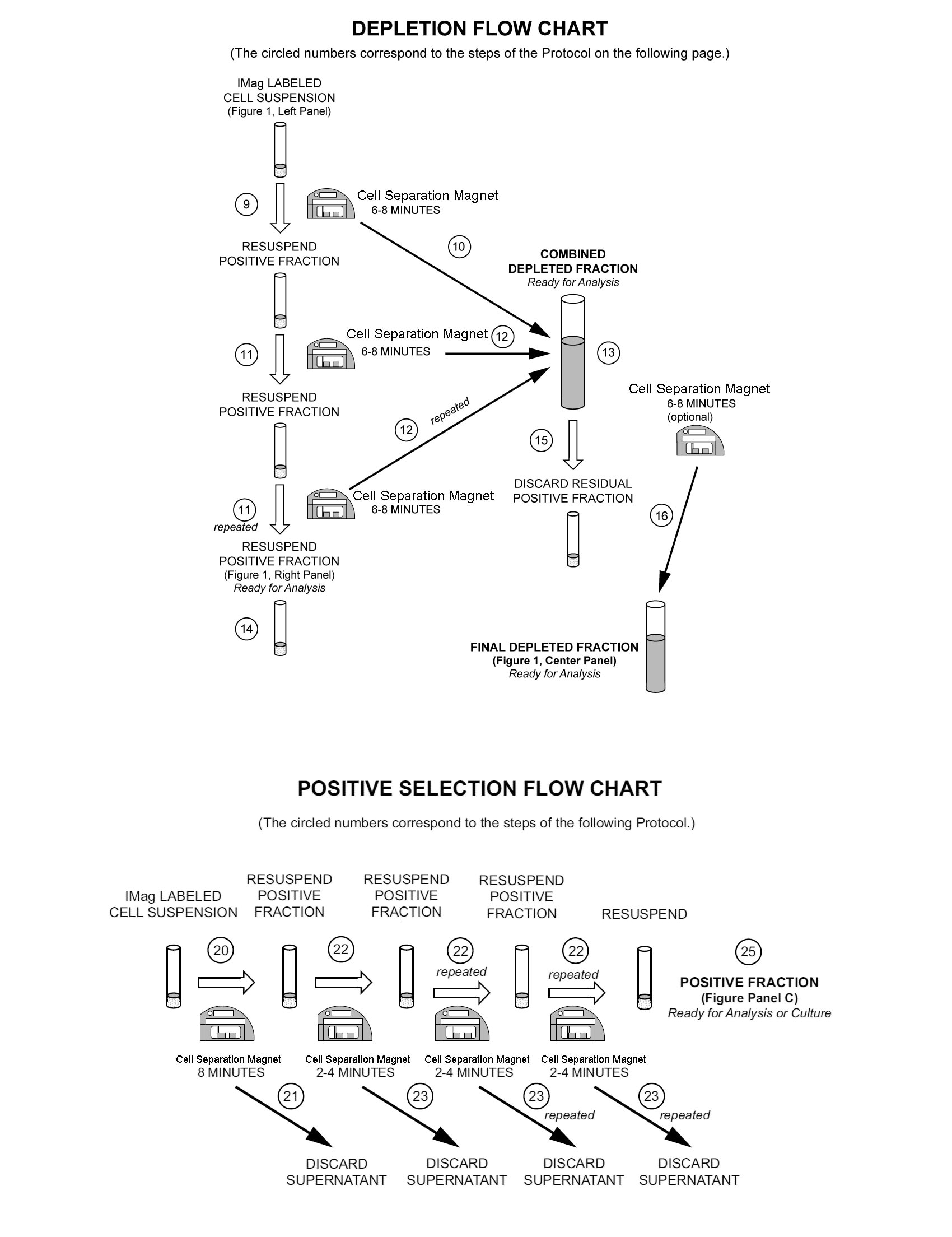
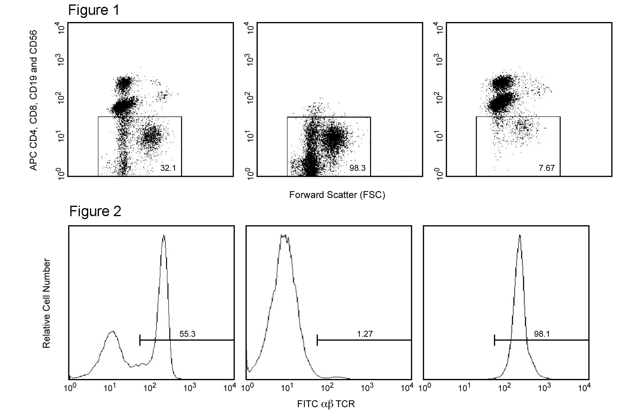
    Figure 1. Depletion of human T and B lymphocytes and NK cells. Human PBMC were stained with APC Mouse Anti-Human CD4 mAb RPA-T4 (Cat. No. 555349), CD8 mAb RPA-T8 (Cat. No. 555369), CD19 mAb HIB19 (Cat. No. 555415), and CD56 mAb B159 (Cat. No. 555518), then labeled with BD IMag™ Anti-Mouse IgG1 Magnetic Particles - DM (Cat. No. 557983). After labeling, the cells were separated using the BD IMag™ Cell Separation Magnet, and the depleted and positive fractions were collected as described in the Magnetic Labeling and Separation Protocol. Please refer to the Depletion Flow Chart to identify the separated cell populations represented in this figure. Unseparated PBMC (left panel), the final depleted fraction (middle panel), and the positive fraction (right panel) were analyzed by flow cytometry. Nonviable cells were eliminated from analysis by staining with Propidium Iodide Staining Solution (Cat. No. 557983), and mononuclear cells were identified by scatter profile. The percentage of non-T-B-NK leukocytes in each sample is given. Flow cytometry was performed on a BD FACSCalibur™ flow cytometry system. Figure 2. Positive selection of rat αß TCR-expressing T lymphocytes. Lewis rat splenocytes were stained with FITC Mouse Anti-Rat αß TCR mAb R73 (Cat. No. 554913), and then labeled with BD IMag™ Anti-Mouse IgG1 Magnetic Particles - DM. After labeling, the cells were separated using the BD IMag™ Cell Separation Magnet, and the negative and positive fractions were collected as described in the Magnetic Labeling and Separation Protocol. Please refer to the Positive Selection Flow Chart to identify the separated cell populations represented in this figure. Unseparated Spleen (left panel), the Negative Fraction (middle panel), and the Positive Fraction (right panel) were analyzed by flow cytometry. Nonviable cells were eliminated from analysis by staining with propidium iodide, and all viable leukocytes are displayed. The percentage of αβ TCR+ T lymphocytes in each sample i

    声明 :本官网所有报价均为常温或者蓝冰运输价格,如有产品需要干冰运输,需另外加收干冰运输费。
    货号:
    557983
    一键复制
    询价
    5mL
    选择数量
    当前规格1件起购 
    配送至
    预计3-4周送达,快递: 免运费,若需干冰额外收费