


Phospho-IKKalpha/beta (Ser176/180) (16A6) Rabbit mAb


 下载产品说明书 下载SDS
 用小程序,查商品更便捷


 收藏
收藏
 对比
对比 咨询
咨询
Product Usage Information
| Application | Dilution |
|---|---|
| Western Blotting | 1:1000 |
| Immunohistochemistry (Paraffin) | 1:75 - 1:300 |
| Flow Cytometry (Fixed/Permeabilized) | 1:400 - 1:1600 |
Specificity/Sensitivity
物种反应性:
人, 小鼠, 大鼠, 猴
参考图片
Immunohistochemical analysis of paraffin-embedded human lung (chronic bronchitis), using Phospho-IKKα/β (Ser176/180) (16A6) Rabbit mAb.免疫组织化学染色分析石蜡包埋人慢性支气管炎组织。所用抗体为Phospho-IKKα/β (Ser176/180) (16A6) Rabbit mAb。
Immunohistochemical analysis of paraffin-embedded human colon carcinoma, showing cytoplasmic localization, using Phospho-IKKα/β (Ser176/180) (16A6) Rabbit mAb.免疫组织化学染色分析石蜡包埋人结肠癌组织,图中所示为细胞质的定位。所用抗体为Phospho-IKKα/β (Ser176/180) (16A6) Rabbit mAb。
Immunohistochemical analysis of paraffin-embedded human breast carcinoma, using Phospho-IKKα/β (Ser176/180) (16A6) Rabbit mAb in the presence of control peptide (left) or Phospho-IKK-alpha/beta (Ser176/180) Blocking Peptide #1023 (right).免疫组织化学染色分析石蜡包埋人乳腺癌组织。所用抗体为Phospho-IKKα/β (Ser176/180) (16A6) Rabbit mAb, 在对照多肽 (左图) 或Phospho-IKK-alpha/beta (Ser176/180) 封闭多肽#1023 (有图)存在下。
Immunohistochemical analysis of paraffin-embedded human colon carcinoma, untreated (left) or λ phosphatase-treated (right), using Phospho-IKKα/β (Ser176/180) (16A6) Rabbit mAb.免疫组织化学染色分析石蜡包埋未经处理 (左图) 和经λ phosphatase处理 (右图) 人结肠癌组织。所用抗体为Phospho-IKKα/β (Ser176/180) (16A6) Rabbit mAb。
Immunohistochemical analysis of paraffin-embedded human gall bladder (chronic cholecystitis), using Phospho-IKKα/β (Ser176/180) (16A6) Rabbit mAb.免疫组织化学染色分析石蜡包埋人胆囊(慢性胆囊炎)组织。所用抗体为Phospho-IKKα/β (Ser176/180) (16A6) Rabbit mAb。
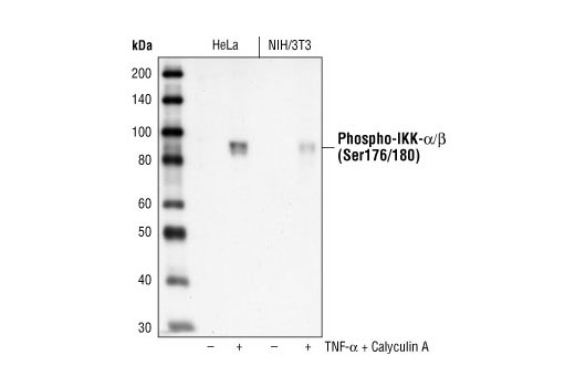
Western blot analysis of extracts from TNF-alpha and Calyculin A treated HeLa and NIH/3T3 cells, using Phospho-IKKα/β (Ser176/180) (16A6) Rabbit mAb.Western免疫印迹。用Phospho-IKKα/β (Ser176/180) (16A6) Rabbit mAb 研究经TNF-alpha 和 Calyculin A处理的HeLa 和 NIH/3T3 细胞的细胞提取液。
Immunohistochemical analysis of frozen H1650 xenograft, showing cytoplasmic localization using Phospho-IKKα/β (Ser176/180)(16A6) Rabbit mAb.免疫组织化学染色研究冰冻H1650 异种移植物,图片显示的是细胞质的定位,所用的抗体为Phospho-IKKα/β (Ser176/180)(16A6) Rabbit mAb。
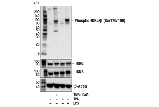
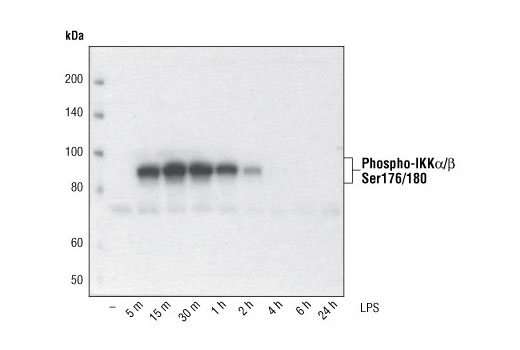
Western blot analysis of extracts from THP-1 cells, differentiated with TPA (#9905, 80 nM for 24h) and treated with 1 μg/ml LPS for the indicated times, using Phospho-IKKα/β (Ser176/180) (16A6) Rabbit mAb.Western免疫印迹。用Phospho-IKKα/β (Ser176/180) (16A6) Rabbit mAb研究用TPA #9905, 80 nM 分化24小时后经1 μg/ml LPS 处理一定时间的 THP-1细胞的细胞提取液。
Flow cytometric analysis of THP-1 cells, untreated (blue) and with TPA and LPS (green) using IKK-α (Ser176/Ser180) phosphate Rabbit mAb. Anti-rabbit IgG (H+L), F(ab')2 Fragment (PE Conjugate) #8885 was used as a secondary antibody.

 危险品化学品经营许可证(不带存储) 许可证编号:沪(杨)应急管危经许[2022]202944(QY)
  营业执照(三证合一)