


1/2


 用小程序,查商品更便捷


产品介绍
产品信息
抗原名称
IL-1beta

来源纯化
通过使用人重组 IL-1β 蛋白对动物进行免疫接种来产生单克隆抗体。
宿主
Rabbit
简单描述
Monoclonal Antibody for studying IL1 beta. Cited in 212 publications. Validated for WB, IF, F. Highly specific and rigorously validated in-house, IL-1β (D3U3E) Rabbit Monoclonal Antibody (CST #12703) is ready to ship.
商品描述
Product Usage Information
| Application | Dilution |
|---|---|
| Western Blotting | 1:1000 |
| Immunofluorescence (Immunocytochemistry) | 1:200 - 1:800 |
| Flow Cytometry (Fixed/Permeabilized) | 1:100 - 1:400 |
分子量
17, 31
研究领域
纤维化,免疫学和肿瘤学,神经科学
应用
反应种属
Human
目标/特异性
Specificity/Sensitivity
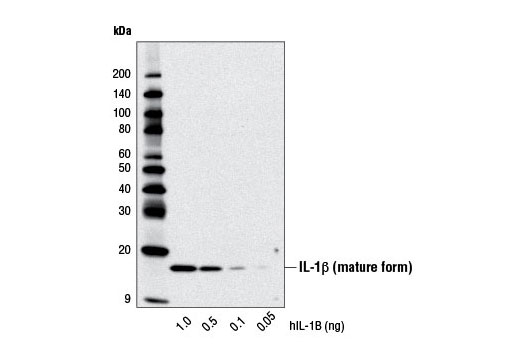
IL-1β (D3U3E) Rabbit mAb 可检测 IL-1β 总蛋白的内源水平。未观察到该抗体可检测成熟 IL-1β 的内源水平。它可检测多达 100 pg 的重组成熟 IL-1β。
物种反应性:
人
敏感性
Endogenous
背景
背景
Interleukin-1β (IL-1β) 是caspase-1 的重要靶标之一,也是一个参与许多免疫和促炎性反应的多功能细胞因子 (1)。它主要由激活的单核细胞和巨噬细胞产生。它通过各种接头蛋白和激酶发出信号,从而激活大量下游靶标 (2-6)。人 IL-1β 可以合成为 31 kDa 前体。为了获得活性,前体必须在 Asp116 和 Ala117 位点之间被 caspase-1 剪切产生 17 kDa 成熟形式 (7,8)。IL-1β 17 kDa 成熟形式的检测是衡量 caspase-1 活性的一个很好指标。
1.Dinarello, C.A. (1998) Int Rev Immunol 16, 457-99.
2.Burns, K. et al. (1998) J Biol Chem 273, 12203-9.
3.Cao, Z. et al. (1996) Nature 383, 443-6.
4.Cao, Z. et al. (1996) Science 271, 1128-31.
5.Wesche, H. et al. (1997) Immunity 7, 837-47.
6.Ninomiya-Tsuji, J. et al. (1999) Nature 398, 252-6.
7.Thornberry, N.A. et al. (1992) Nature 356, 768-74.
8.Cerretti, D.P. et al. (1992) Science 256, 97-100.
研究领域
纤维化,免疫学和肿瘤学,神经科学
翻译后修饰
Unmodified
制备和贮存
保存方式
Supplied in 10 mM sodium HEPES (pH 7.5), 150 mM NaCl, 100 µg/ml BSA, 50% glycerol and less than 0.02% sodium azide. Store at –20°C. Do not aliquot the antibody.For a carrier free (BSA and azide free) version of this product see product #18689.
数据库链接
Entrez-Gene ID
3553
UniProt ID
P01584
参考图片
Flow cytometric analysis of THP-1 cells, untreated (blue) or LPS-treated (100 ng/ml, 3 hr; green), using IL-1β (D3U3E) Rabbit mAb. Anti-rabbit IgG (H+L), F(ab')2 Fragment (Alexa Fluor® 647 Conjugate) #4414 was used as a secondary antibody.
Confocal immunofluorescent analysis of THP-1 cells, untreated (left) or LPS-treated (500 ng/ml, 2 hr; right), using IL-1β (D3U3E) Rabbit mAb (green). Actin filaments were labeled with DY-554 phalloidin (red).
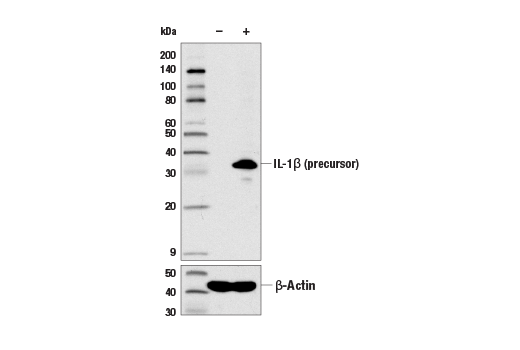
Western blot analysis of extracts from THP-1 cells, untreated (-) or LPS-treated (100 ng/ml, 3 hr; +), using IL-1β (D3U3E) Rabbit mAb (upper) and β-Actin (D6A8) Rabbit mAb #8457 (lower).
Western blot analysis of recombinant Human Interleukin-1β (hIL-1β) #8900 using IL-1β (D3U3E) Rabbit mAb.
声明 :本官网所有报价均为常温或者蓝冰运输价格,如有产品需要干冰运输,需另外加收干冰运输费。

 危险品化学品经营许可证(不带存储) 许可证编号:沪(杨)应急管危经许[2022]202944(QY)
  营业执照(三证合一)