


BD Horizon™ BV480 Rat Anti-Mouse CD25


 下载产品说明书
 用小程序,查商品更便捷


 收藏
收藏
 对比
对比 咨询
咨询

参考图片
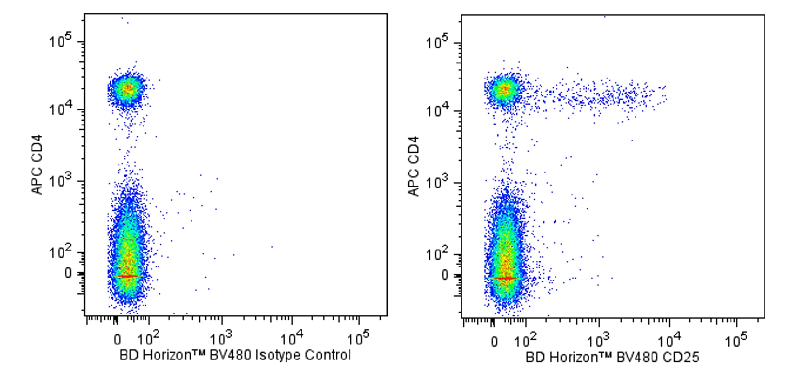
Two-color flow cytometric analysis of CD25 expression on mouse splenocytes. Mouse splenic leucocytes were preincubated with Purified Rat Anti-Mouse CD16/CD32 antibody (Mouse BD Fc Block™) (Cat. No. 553141/553142). The cells were then stained with APC Rat Anti-Mouse CD4 antibody (Cat. No. 553051/561091) and either BD Horizon™ BV480 Rat IgG1, λ Isotype Control (Cat. No. 566128; Left Plot) or BD Horizon BV480 Rat Anti-Mouse CD25 antibody (Cat. No. 566202/566120; Right Plot). Two-color flow cytometric dot plots showing the correlated expression of CD25 (or Ig Isotype control staining) versus CD4 were derived from gated events with the forward and side light-scatter characteristics of viable splenocytes. Flow cytometric analysis was performed using a BD LSRFortessa™ Cell Analyzer System.
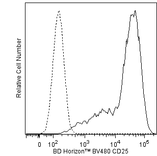
Flow cytometric analysis of CD25 expression on activated mouse splenocytes. Mouse splenic leucocytes were stimulated for 3 days with concanavalin A (ConA). The cells were preincubated with Purified Rat Anti-Mouse CD16/CD32 antibody and then stained with and either BD Horizon BV480 Rat IgG1, λ Isotype Control (dashed line histogram) or BD Horizon BV480 Rat Anti-Mouse CD25 antibody (solid line histogram). The fluorescence histogram showing CD25 expression (or Ig Isotype control staining) was derived from events with the forward and side light-scatter characteristics of viable lymphoblasts. Flow cytometric analysis was performed using a BD LSRFortessa™ Cell Analyzer System.
Two-color flow cytometric analysis of CD25 expression on mouse splenocytes. Mouse splenic leucocytes were preincubated with Purified Rat Anti-Mouse CD16/CD32 antibody (Mouse BD Fc Block™) (Cat. No. 553141/553142). The cells were then stained with APC Rat Anti-Mouse CD4 antibody (Cat. No. 553051/561091) and either BD Horizon™ BV480 Rat IgG1, λ Isotype Control (Cat. No. 566128; Left Plot) or BD Horizon BV480 Rat Anti-Mouse CD25 antibody (Cat. No. 566202/566120; Right Plot). Two-color flow cytometric dot plots showing the correlated expression of CD25 (or Ig Isotype control staining) versus CD4 were derived from gated events with the forward and side light-scatter characteristics of viable splenocytes. Flow cytometric analysis was performed using a BD LSRFortessa™ Cell Analyzer System.
Flow cytometric analysis of CD25 expression on activated mouse splenocytes. Mouse splenic leucocytes were stimulated for 3 days with concanavalin A (ConA). The cells were preincubated with Purified Rat Anti-Mouse CD16/CD32 antibody and then stained with and either BD Horizon BV480 Rat IgG1, λ Isotype Control (dashed line histogram) or BD Horizon BV480 Rat Anti-Mouse CD25 antibody (solid line histogram). The fluorescence histogram showing CD25 expression (or Ig Isotype control staining) was derived from events with the forward and side light-scatter characteristics of viable lymphoblasts. Flow cytometric analysis was performed using a BD LSRFortessa™ Cell Analyzer System.

 危险品化学品经营许可证(不带存储) 许可证编号:沪(杨)应急管危经许[2022]202944(QY)
  营业执照(三证合一)