


1/2
BD Horizon™ PE-CF594 Rat Anti-Mouse CD49b


品牌: BD Pharmingen
 下载产品说明书 下载SDS
 用小程序,查商品更便捷


 收藏
收藏
 对比
对比 咨询
咨询反应种属:
Mouse (QC Testing)
Mouse (QC Testing)
来源宿主:
Rat LEW, also known as Lewis IgM, κ
Rat LEW, also known as Lewis IgM, κ
产品介绍
产品信息
耦联标记
PE-CF594

抗原名称
CD49b (Integrin α2)
宿主
Rat LEW, also known as Lewis IgM, κ
免疫原
Mouse (C57BL/6) NK1.1+ cells propagated with rIL-2
简单描述
The rat anti-mouse CD49b monoclonal antibody (clone DX5) specifically binds to the integrin α2 chain (CD49b). CD49b is a 150 kDa transmembrane glycoprotein that non-covalently associates with CD29 (integrin β1) to form the integrin α2β1 complex known as VLA-2. The rat anti-mouse CD49b antibody (clone DX5) has been reported to identify the majority of NK cells and a small T-cell subpopulation in most mouse strains (e.g., A/J, AKR, BALB/c, C3H/HeJ, C57BL/6, C57BL/10, C57BR, C58, CBA/Ca, DBA/1, DBA/2, SJL, SWR, 129/J, but not NOD). The DX5 antibody also recognizes platelets that express high levels of CD49b. Multiparameter flow cytometric analysis has demonstrated that most lymphocytes which express NK-1.1 (NKR-P1B and NKR-P1C), as detectable by mouse anti-mouse NK-1.1 antibody (clone PK136), also express the DX5 antigen. Small DX5+ NK-1.1- and DX5- NK-1.1+ cell subsets are found, especially among the CD3-positive cell population. Some CD49b+ NK cells have been reported to gradually lose reactivity with the rat anti-mouse CD49b antibody (clone DX5) when cultured in the presence of recombinant human IL-2. The resulting DX5-negative cells have weakened cytotoxic activity when compared to the remaining DX5+ cells. This indicates that the DX5 antibody distinguishes functional subsets of NK cells. No activation or blocking activity of the rat anti-mouse antibody (clone DX5) has been observed. Staining of splenic NK cells with this antibody reportedly can be blocked by hamster anti-mouse CD49b antibody (clone HMα2).
This antibody is conjugated to BD Horizon™ PE-CF594, which has been developed exclusively by BD Biosciences as a better alternative to PE-Texas Red. PE-CF594 excites and emits at similar wavelengths to PE-Texas Red yet exhibits improved brightness and spectral characteristics. Due to PE having maximal absorption peaks at 496 nm and 564 nm, PE-CF594 can be excited by the blue (488-nm), green (532-nm) and yellow-green (561-nm) lasers and can be detected with the same filter set as PE-Texas Red (eg 610/20-nm filter).
商品描述
DX5
The rat anti-mouse CD49b monoclonal antibody (clone DX5) specifically binds to the integrin α2 chain (CD49b). CD49b is a 150 kDa transmembrane glycoprotein that non-covalently associates with CD29 (integrin β1) to form the integrin α2β1 complex known as VLA-2. The rat anti-mouse CD49b antibody (clone DX5) has been reported to identify the majority of NK cells and a small T-cell subpopulation in most mouse strains (e.g., A/J, AKR, BALB/c, C3H/HeJ, C57BL/6, C57BL/10, C57BR, C58, CBA/Ca, DBA/1, DBA/2, SJL, SWR, 129/J, but not NOD). The DX5 antibody also recognizes platelets that express high levels of CD49b. Multiparameter flow cytometric analysis has demonstrated that most lymphocytes which express NK-1.1 (NKR-P1B and NKR-P1C), as detectable by mouse anti-mouse NK-1.1 antibody (clone PK136), also express the DX5 antigen. Small DX5+ NK-1.1- and DX5- NK-1.1+ cell subsets are found, especially among the CD3-positive cell population. Some CD49b+ NK cells have been reported to gradually lose reactivity with the rat anti-mouse CD49b antibody (clone DX5) when cultured in the presence of recombinant human IL-2. The resulting DX5-negative cells have weakened cytotoxic activity when compared to the remaining DX5+ cells. This indicates that the DX5 antibody distinguishes functional subsets of NK cells. No activation or blocking activity of the rat anti-mouse antibody (clone DX5) has been observed. Staining of splenic NK cells with this antibody reportedly can be blocked by hamster anti-mouse CD49b antibody (clone HMα2).
This antibody is conjugated to BD Horizon™ PE-CF594, which has been developed exclusively by BD Biosciences as a better alternative to PE-Texas Red. PE-CF594 excites and emits at similar wavelengths to PE-Texas Red yet exhibits improved brightness and spectral characteristics. Due to PE having maximal absorption peaks at 496 nm and 564 nm, PE-CF594 can be excited by the blue (488-nm), green (532-nm) and yellow-green (561-nm) lasers and can be detected with the same filter set as PE-Texas Red (eg 610/20-nm filter).
同种型
Rat LEW, also known as Lewis IgM, κ
克隆号
克隆 DX5 (RUO)
浓度
0.2 mg/ml
产品详情
PE-CF594
BD Horizon™ PE-CF594 dye is a part of the BD PE family of dyes. This tandem fluorochrome is comprised of a R-Phycoerythrin (PE) donor that has excitation maxima (Ex Max) of 496-nm and 566-nm and an acceptor dye with an emission maximum (Em Max) at 615-nm. PE-CF594, driven by BD innovation, is designed to be excited by the blue (488-nm), Green (532-nm) and yellow-green (561-nm) lasers and detected using an optical filter centered near 615 nm (e.g., a 610/20-nm bandpass filter). The donor dye can be excited by the Blue (488-nm), Green (532-nm) and yellow-green (561-nm) lasers and the acceptor dye can be excited by the green (532-nm) laser resulting in cross-laser excitation and fluorescence spillover. Please ensure that your instrument’s configurations (lasers and optical filters) are appropriate for this dye.

PE-CF594
Yellow-Green 488 nm, 532 nm, 561 nm
496 nm, 566 nm
615 nm
应用
实验应用
Flow cytometry (Routinely Tested)
反应种属
Mouse (QC Testing)
目标/特异性
CD49b (Integrin α2)
背景
别名
itga2; Integrin alpha-2; DX5; Pan NK cell marker; VLAA2; VLA-2 alpha chain
制备和贮存
存储溶液
Aqueous buffered solution containing BSA, protein stabilizer, and ≤0.09% sodium azide.
保存方式
Aqueous buffered solution containing BSA, protein stabilizer, and ≤0.09% sodium azide.
文献
文献
研发参考(3)
1. Arase H, Saito T, Phillips JH, Lanier LL. Cutting edge: the mouse NK cell-associated antigen recognized by DX5 monoclonal antibody is CD49b (alpha 2 integrin, very late antigen-2). J Immunol. 2001; 167(3):1141-1144. (Clone-specific: Blocking, Cytotoxicity, Flow cytometry).
2. Moore TA, von Freeden-Jeffry U, Murray R, Zlotnik A. Inhibition of gamma delta T cell development and early thymocyte maturation in IL-7 -/- mice. J Immunol. 1996; 157(6):2366-2373. (Biology).
3. Ortaldo JR, Winkler-Pickett R, Mason AT, Mason LH. The Ly-49 family: regulation of cytotoxicity and cytokine production in murine CD3+ cells. J Immunol. 1998; 160(1):1158-1165. (Biology).
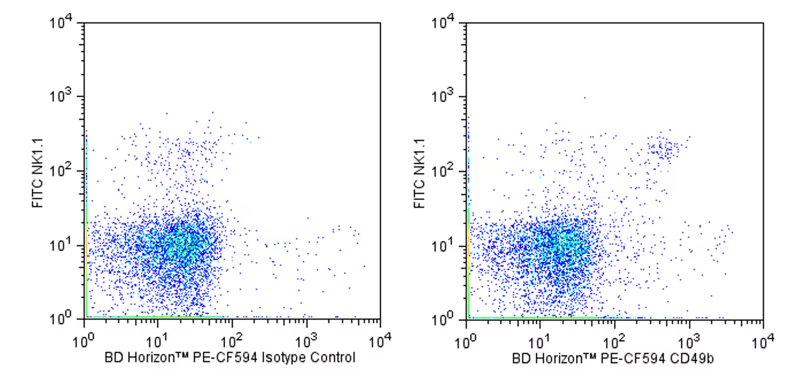
参考图片
Multicolor flow cytometric analysis of CD49b expressed on mouse splenocytes. Splenocytes from C57BL/6 mice were stained with a FITC Mouse Anti-Mouse NK1.1 antibody (Cat. No. 553857/561067) and with either BD Horizon™ PE-CF594 Rat IgM, κ Isotype Control (Cat. No. 562489, Left Panel) or BD Horizon™ PE-CF594 Rat Anti-Mouse CD49b antibody (Cat. No. 562453, Right Panel). Two color flow cytometric dot plots showing the expression of NK1.1 versus CD49b (or Ig isotype control staining) were derived from gated events with the forward and side light-scatter characteristics of viable splenocytes. Flow cytometry was performed using a BD™ LSR II Flow Cytometer System.
声明 :本官网所有报价均为常温或者蓝冰运输价格,如有产品需要干冰运输,需另外加收干冰运输费。

 危险品化学品经营许可证(不带存储) 许可证编号:沪(杨)应急管危经许[2022]202944(QY)
  营业执照(三证合一)