


 下载产品说明书
 用小程序,查商品更便捷


 收藏
收藏
 对比
对比 咨询
咨询
Specificity/Sensitivity
参考图片
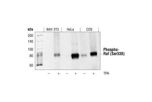
Western blot analysis of extracts from NIH/3T3, HeLa and COS cells, untreated or treated with TPA, using Phospho-c-Raf (Ser338) (56A6) Rabbit mAb #9427.
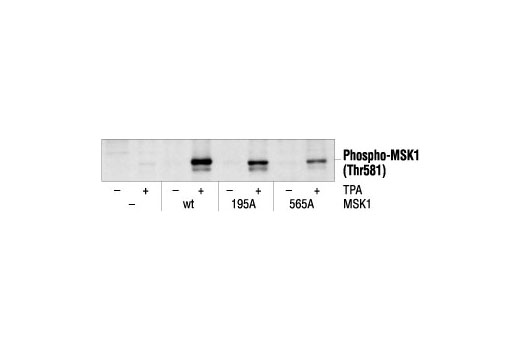
Western blot analysis of extracts from 293 cells, transfected with wt MSK1 or mutant MSK1, untreated or TPA-treated (200 nM), using Phospho-MSK1 (Thr581) Antibody.
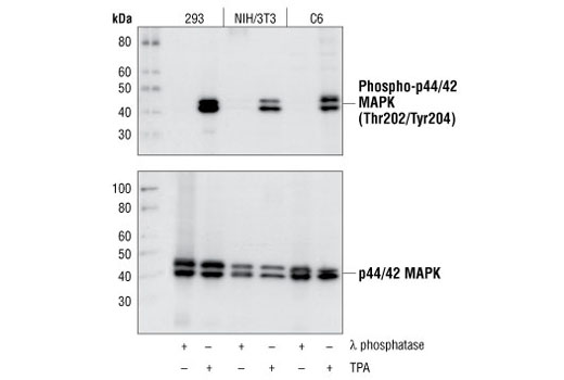
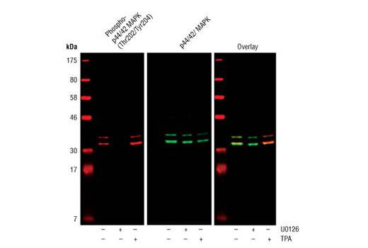
Western blot analysis of extracts from 293, NIH/3T3 and C6 cells, treated with λ phosphatase or TPA #4174 as indicated, using Phospho-p44/42 MAPK (Erk1/2) (Thr202/Tyr204) (D13.14.4E) XP® Rabbit mAb (upper), or p44/42 MAPK (Erk1/2) (137F5) Rabbit mAb #4695 (lower).
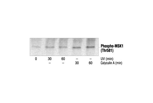
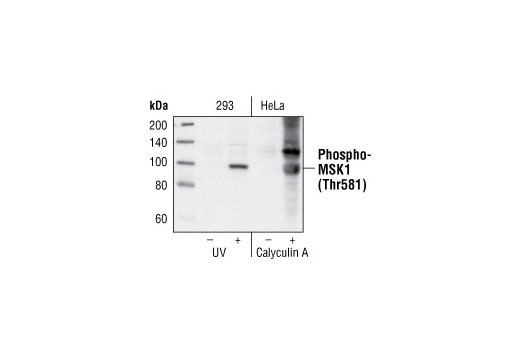
Western blot analysis of extracts from 293 cells untreated or treated with UV and HeLa cells untreated or treated with calyculin A, using Phospho-MSK1 (Thr581) Antibody.
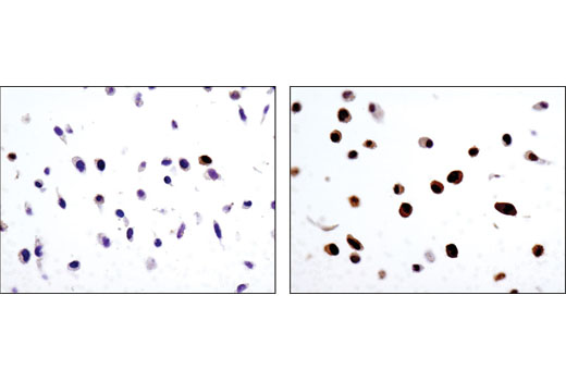
Immunohistochemical analysis using Phospho-p44/42 MAPK (Erk1/2) (Thr202/Tyr204) (D13.14.4E) XP® Rabbit mAb on SignalSlide™ Phospho-p44/42 MAPK (Thr202/Tyr204) IHC Controls #8103 (paraffin-embedded NIH/3T3 cells, treated with U0126 #9903 (left) or TPA #4174 (right).
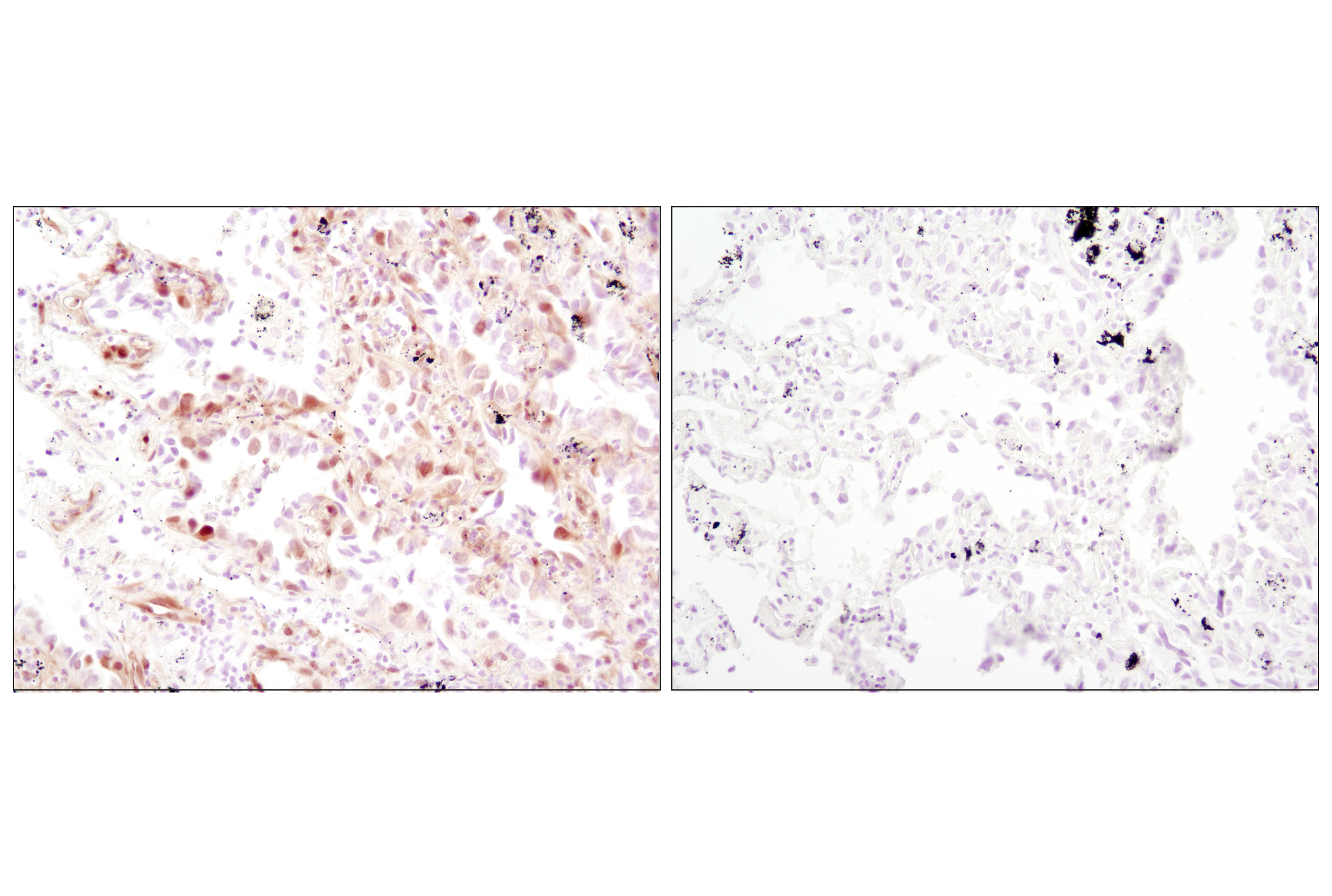
Immunohistochemical analysis of paraffin-embedded human lung carcinoma, untreated (left) or λ phosphatase-treated (right), using Phospho-p44/42 MAPK (Erk1/2) (Thr202/Tyr204) (D13.14.4E) XP® Rabbit mAb.
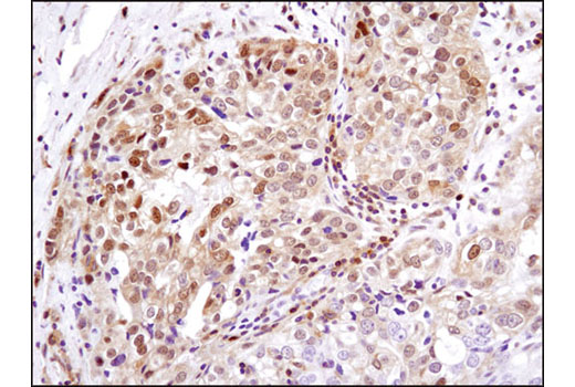
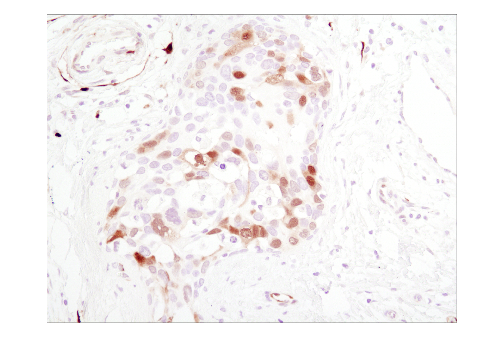
Immunohistochemical analysis of paraffin-embedded human breast carcinoma using Phospho-p44/42 MAPK (Erk1/2) (Thr202/Tyr204) (D13.14.4E) XP® Rabbit mAb.
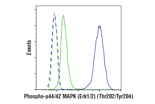
Flow cytometric analysis of Jurkat cells, U0126-treated (blue) or PMA-treated (green), using Phospho-p44/42 MAPK (Erk1/2) (Thr202/Tyr204) (D13.14.4E) XP® Rabbit mAb.
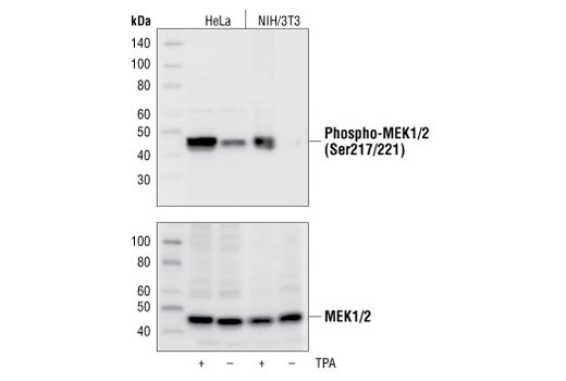
Western blot analysis of extracts from untreated or TPA-treated HeLa and NIH/3T3 cells, using Phospho-MEK1/2 (Ser217/221) (41G9) Rabbit mAb #9154 (upper), or MEK1/2 Antibody #9122 (lower).
Immunohistochemical analysis of paraffin-embedded human breast carcinoma, showing nuclear localization, using Phospho-MSK1 (Thr581) Antibody.
Flow cytometric analysis of Jurkat cells, U0126-treated (blue) or PMA-treated (green), using Phospho-p44/42 MAPK (Thr202/Tyr204) (D13.14.4E) XP® Rabbit mAb #4370.
Confocal immunofluorescent analysis of C2C12 cells, U0126-treated (left, #9903 10 μM for 1 h) or TPA-treated (right, #9905 200 nM for 15 min), using Phospho-p44/42 MAPK (Thr202/Tyr204) (D13.14.4E) Rabbit mAb #4370 (green). Actin filaments have been labeled with Alexa Fluor® 555 phalloidin (red). Blue pseudocolor = DRAQ5™ (fluorescent DNA dye).
Immunohistochemical analysis of paraffin-embedded human lung carcinoma, untreated (left) or λ phosphatase-treated (right), using Phospho-p44/42 MAPK (Thr202/Tyr204) (D13.14.4E) Rabbit mAb #4370.
Western blot analysis of extracts from 293, NIH/3T3 and C6 cells, treated with λ phosphatase or TPA as indicated, using Phospho-p44/42 MAPK (Thr202/Tyr204) (D13.14.4E) Rabbit mAb #4370 (upper), or p44/42 MAP Kinase (137F5) Rabbit mAb #4695 (lower).
Western blot analysis of extracts from 293 cells untreated or treated with UV and HeLa cells untreated or treated with calyculin A using Phospho-MSK1 (Thr581) Antibody #9595.
Western blot analysis of extracts from NIH3T3, HeLa and COS cells, untreated or treated with TPA, using Phospho-c-Raf (Ser338) (56A6) Rabbit mAb.
Western blot analysis of extracts from untreated or TPA-treated 293 and NIH/3T3 cells using Phospho-p90RSK (Ser380) (9D9) Rabbit mAb #9335.
Western blot analysis of extracts from COS cells, untreated or treated with either U0126 #9903 (10 µM for 1h) or TPA #4174 (200 nM for 10 m), using Phospho-p44/42 MAPK (Erk1/2) (Thr202/Tyr204) (D13.14.4E) XP® Rabbit mAb #4370 and p44/42 MAPK (Erk1/2) (3A7) Mouse mAb #9107.
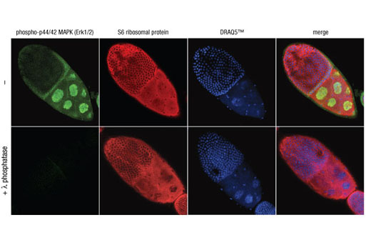
Confocal immunofluorescent analysis of Drosophila egg chambers, untreated (top) or λ phosphatase-treated (bottom), using Phospho-p44/42 MAPK (Erk1/2) (Thr202/Tyr204) (D13.14.4E) XP® Rabbit mAb #4370 (green) and S6 Ribosomal Protein (54D2) Mouse mAb #2317 (red). Blue pseudocolor = DRAQ5® #4084 (fluorescent DNA dye).
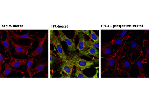
Confocal immunofluorescent analysis of NIH/3T3 cells, serum-starved (left), treated with TPA #4174 (200 nM, 15 min; center), or treated with TPA followed by λ phosphatase (right), using Phospho-p90RSK (Ser380) (D3H11) Rabbit mAb (green). Actin filaments were labeled with DY-554 phalloidin (red). Blue pseudocolor = DRAQ5® #4084 (fluorescent DNA dye).
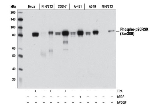
Western blot analysis of extracts from various cell lines, starved overnight and either untreated (-) or treated (+) with TPA #4174 (200 nM, 15 min), Human Epidermal Growth Factor (hEGF) #8916 (100 ng/mL, 15 min), or Human Platelet-Derived Growth Factor BB (hPDGF-BB) #8912 (100 ng/mL, 15 min) as indicated, using Phospho-p90RSK (Ser380) (D3H11) Rabbit mAb.
Western blot analysis of extracts from 293 cells, untreated, UV-treated or calyculin A-treated, using Phospho-MSK1 (Thr581) Antibody.
After the primary antibody is bound to the target protein, a complex with HRP-linked secondary antibody is formed. The LumiGLO* is added and emits light during enzyme catalyzed decomposition.
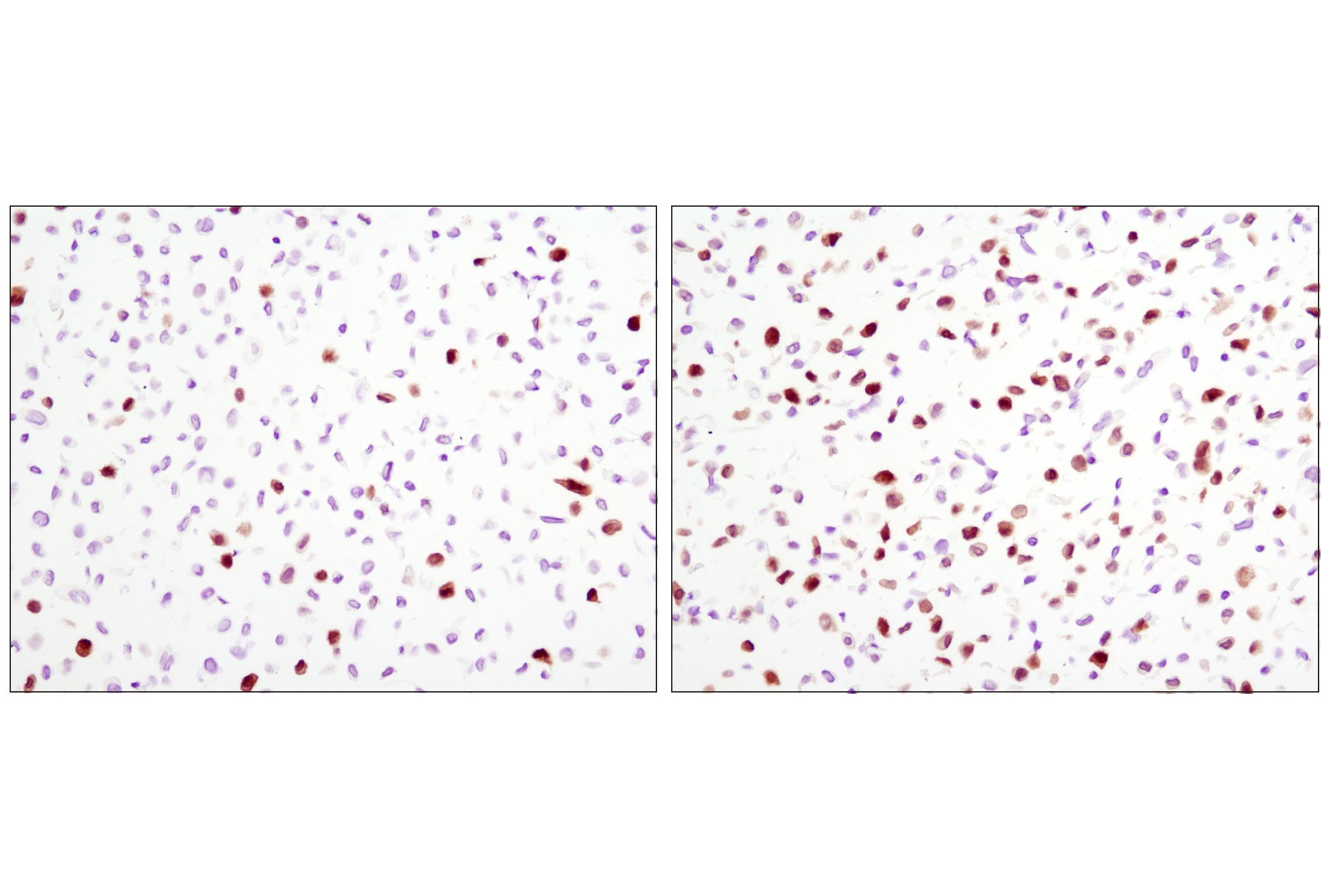
Immunohistochemical analysis of paraffin-embedded HeLa cell pellets, untreated (left) or treated with TPA #4174 (right), using Phospho-p90RSK (Ser380) (D3H11) Rabbit mAb.
Immunohistochemical analysis of paraffin-embedded human ovarian carcinoma using Phospho-p90RSK (Ser380) (D3H11) Rabbit mAb.
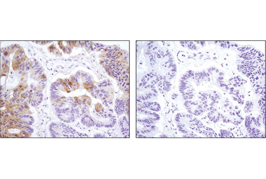
Immunohistochemical analysis of paraffin-embedded human colon carcinoma, control (left) or λ phosphatase-treated (right), using Phospho-p90RSK (Ser380) (D3H11) Rabbit mAb.
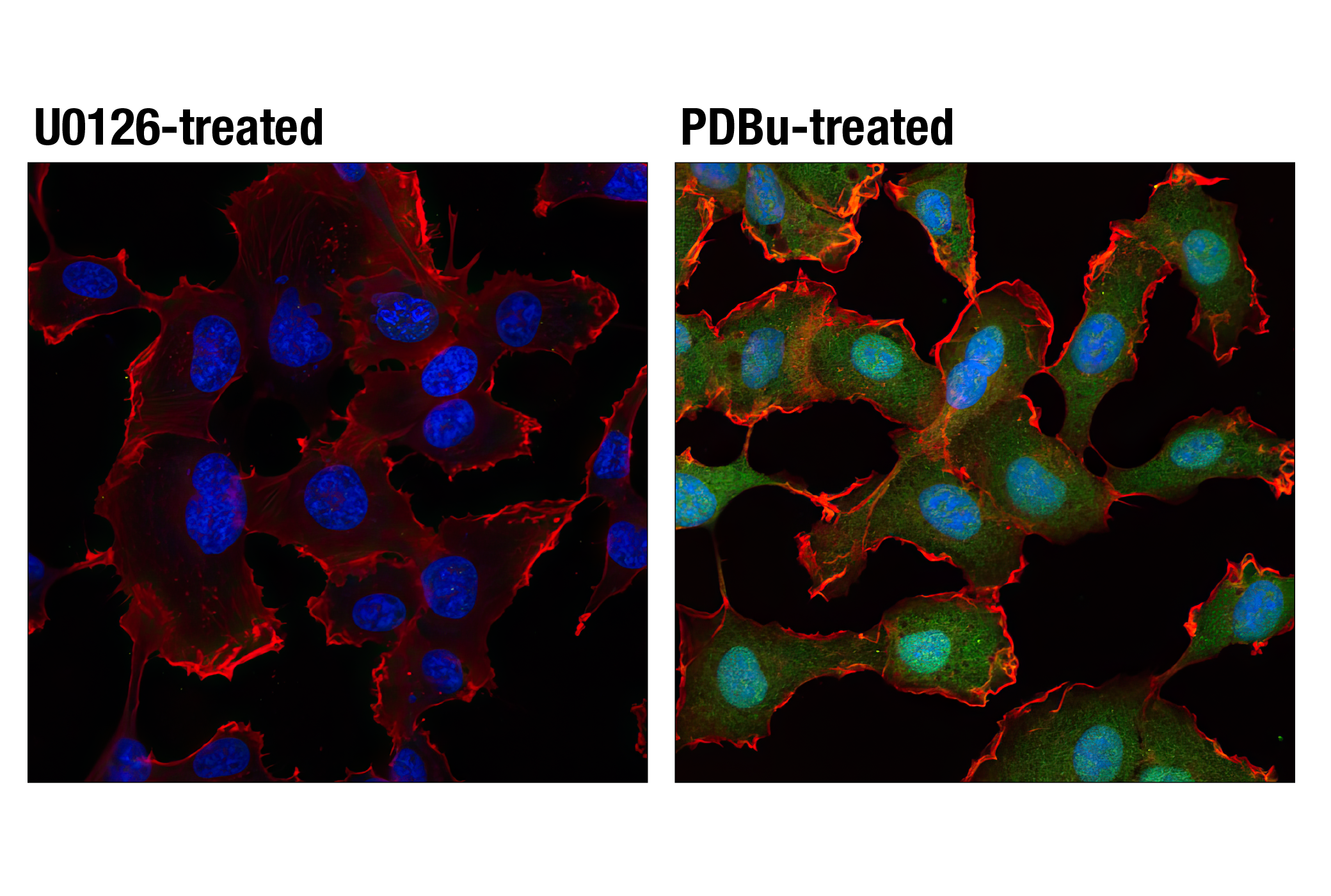
Confocal immunofluorescent analysis of HT1080 cells, starved overnight then treated with U0126 #9903 (10 uM, 2 h; left) or PDBu (Phorbol 12,13-Dibutyrate) #12808 (100 nM, 15 m; right) using Phospho-p44/42 MAPK (Erk1/2) (Thr202/Tyr204) (D13.14.4E) XP® Rabbit mAb #4370 (green) and β-Actin (8H10D10) Mouse mAb #3700 (red). Blue pseudocolor = DRAQ5® #4084 (fluorescent DNA dye).
Western blot analysis of extracts from untreated or TPA-treated HeLa and NIH/3T3 cells, using Phospho-MEK1/2 (Ser217/221) (41G9) Rabbit mAb (upper), or MEK1/2 Antibody #9122 (lower).

 危险品化学品经营许可证(不带存储) 许可证编号:沪(杨)应急管危经许[2022]202944(QY)
  营业执照(三证合一)