


 下载产品说明书
 用小程序,查商品更便捷


 收藏
收藏
 对比
对比 咨询
咨询
Specificity/Sensitivity
参考图片
After the primary antibody is bound to the target protein, a complex with HRP-linked secondary antibody is formed. The LumiGLO* is added and emits light during enzyme catalyzed decomposition.
After the primary antibody is bound to the target protein, a complex with HRP-linked secondary antibody is formed. The LumiGLO* is added and emits light during enzyme catalyzed decomposition.
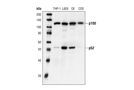
Western blot analysis of extracts from THP-1, L929, C6, and COS cells, using NF-kappaB2 p100 Antibody.
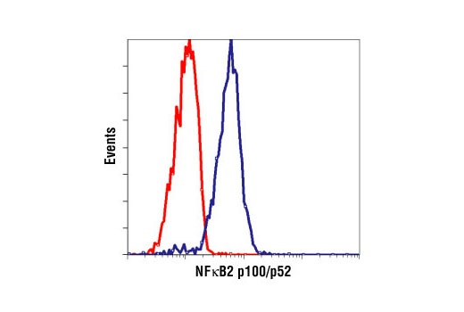
Flow cytometric analysis of HeLa cells, using NF-kappaB2 p100/p52 (18D10) Rabbit mAb (Human Specific) (blue) compared to a nonspecific negative control antibody (red).
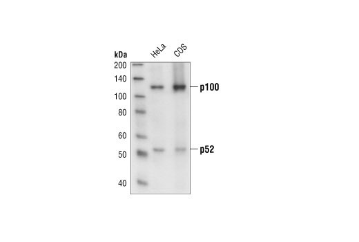
Western blot analysis of extracts from HeLa, and COS cells, using NF-kB2 p100/p52 (18D10) Rabbit mAb (Human Specific).
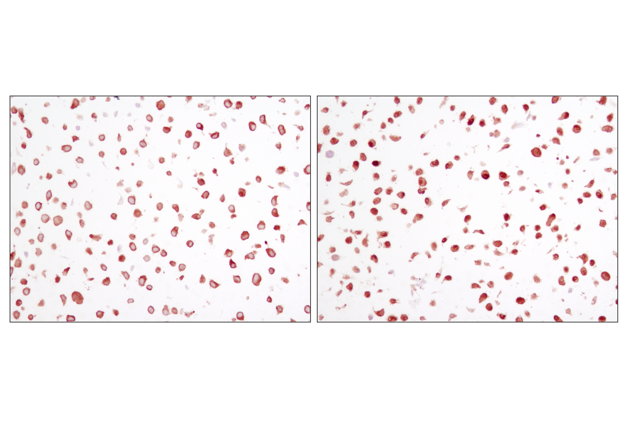
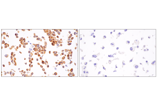
Immunohistochemical analysis of paraffin-embedded human osteosarcoma, using NF-kappaB2 p100/p52 (18D10) Rabbit mAb (Human Specific).
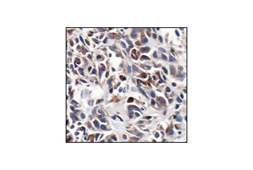
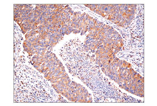
Immunohistochemical analysis of paraffin-embedded human lung carcinoma, using NF-kappaB2 p100/p52 (18D10) Rabbit mAb (Human Specific).
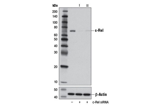
Western blot analysis of extracts from K562, Raji and 293 cells, using c-Rel Antibody.
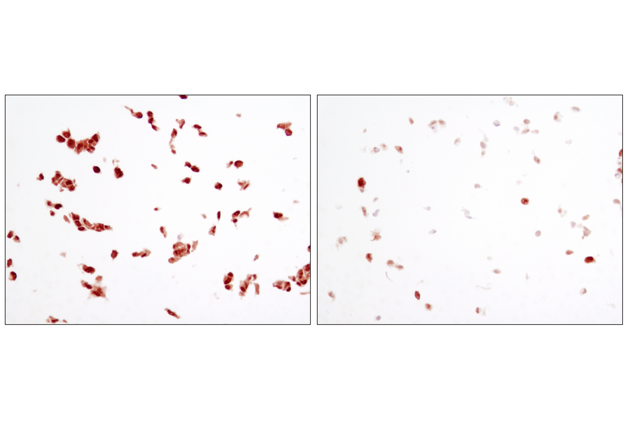
Immunohistochemical analysis of paraffin-embedded human breast carcinoma using c-Rel Antibody in the presence of control peptide (left) or antigen-specific peptide (right).
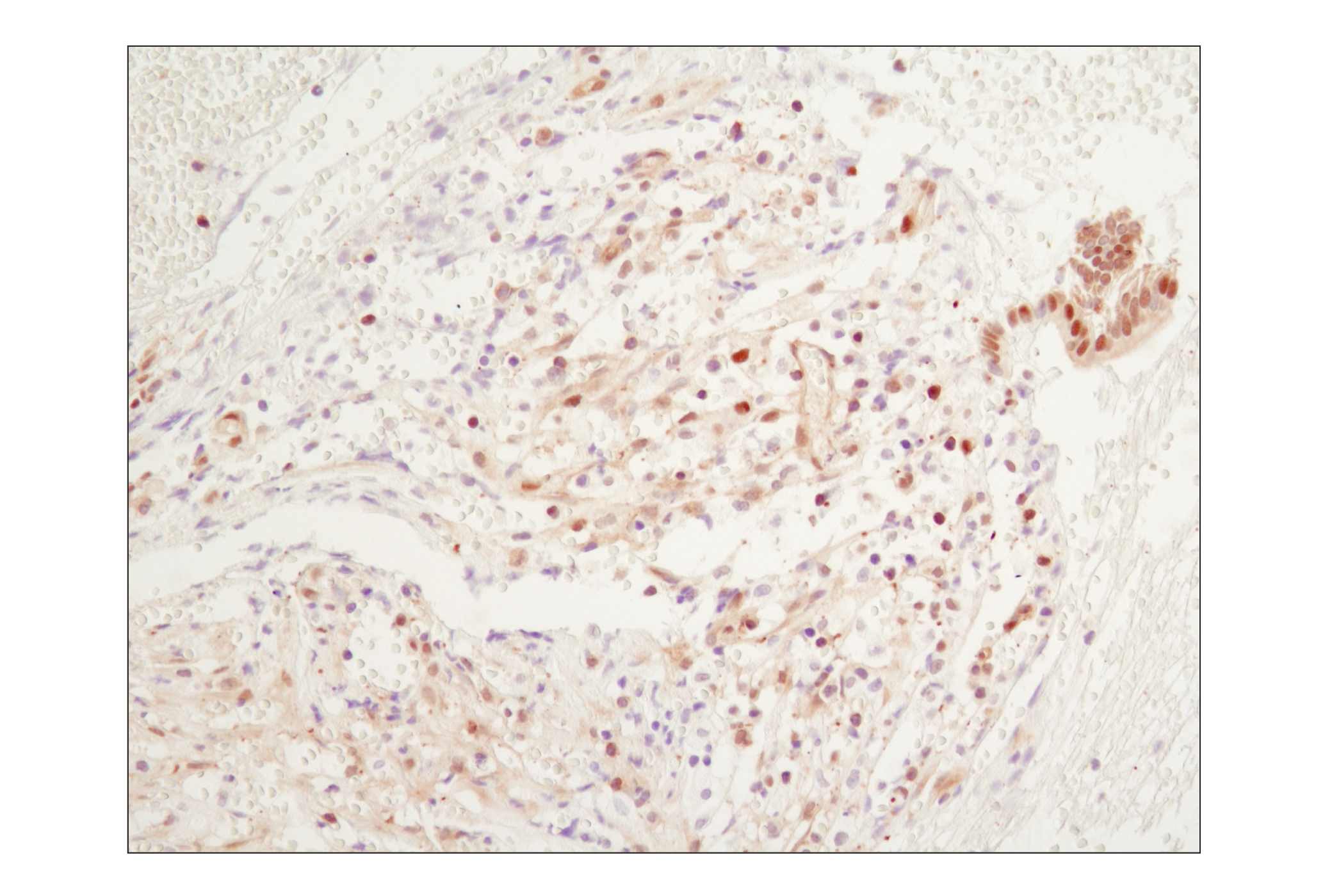
Immunohistochemical analysis of paraffin-embedded Hodgkin's lymphoma, showing nuclear and cytoplasmic localization using c-Rel Antibody.
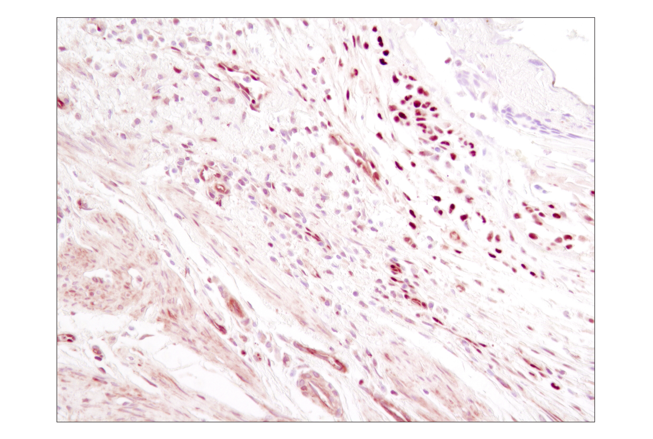
Immunohistochemical analysis of paraffin-embedded human skin, using c-Rel Antibody.
Flow cytometric analysis of K562 cells using c-Rel Antibody (blue) compared to a nonspecific negative control antibody (red).
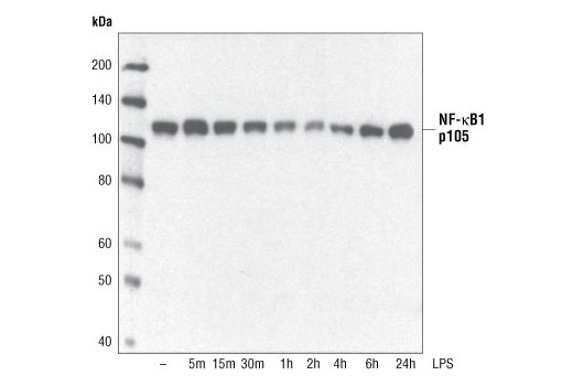
Western blot analysis of extracts from THP-1 cells, differentiated with TPA (#9905, 80 nM for 24 h) and treated with 1 μg/ml LPS for the indicated times, using NF-κB1 p105 Antibody.
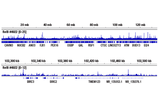
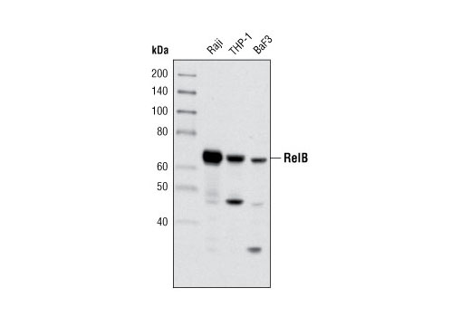
Western blot analysis of extracts from Raji, THP-1 and BaF3 cells using RelB (C1E4) Rabbit mAb.
Western blot analysis of extracts from Raji, THP-1 and BaF3 cells using RelB (C1E4) Rabbit mAb #4922.Western免疫印迹。用RelB (C1E4) Rabbit mAb #4922研究Raji, THP-1和BaF3 细胞的细胞提取液。
Western blot analysis of extracts from K562, Raji and 293 cells using c-Rel Antibody #4727.Western免疫印迹。用 c-Rel Antibody #4727研究 K562, Raji 和293 细胞的细胞提取液。
Western blot analysis of extracts from HeLa cells, untreated or treated with TNF-α (12 ng/ml) for the indicated amounts of time, using NF-kB p105/p50 Antibody #3035.Western免疫印迹。用 NF-kB p105/p50 Antibody #3035研究未经处理的和经TNF-α (12 ng/ml) 处理的一定时间的HeLa细胞的细胞提取液。
Western blot analysis of extracts from various cell types using NF-kB2 p100/p52 Antibody #4882.Western免疫印迹。用NF-kB2 p100/p52 Antibody #4882研究各种细胞类型的细胞提取液。
Western blot analysis of extracts from HeLa and COS cells using NF-kB2 p100/p52 (18D10) Rabbit mAb (Human Specific) #3017.Western免疫印迹。用NF-kB2 p100/p52 (18D10) Rabbit mAb (Human Specific) #3017研究 HeLa 和COS 细胞的细胞提取液。
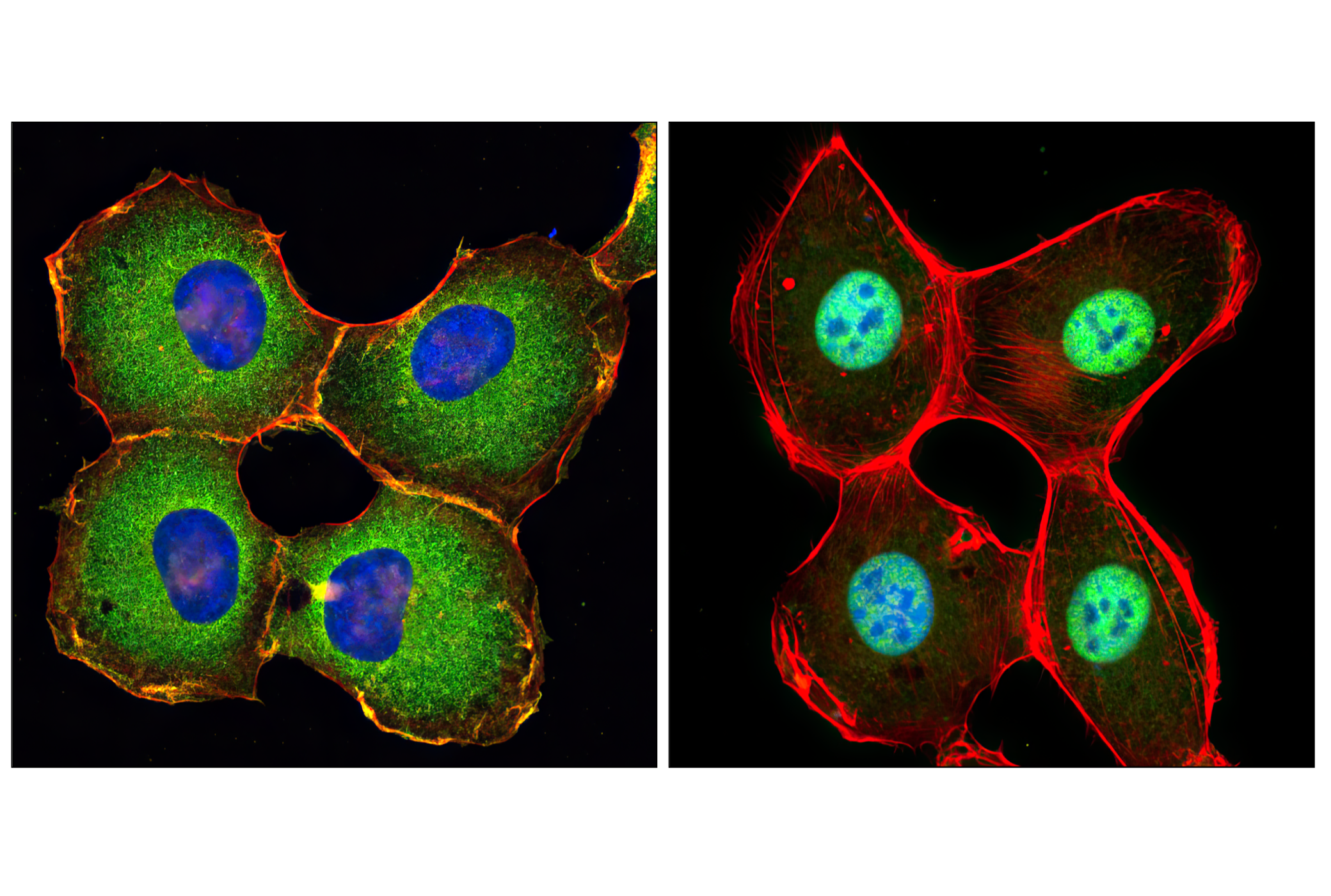
Confocal immunofluorescent analysis of HeLa cells, untreated (left) or TNFα-treated (#2169, 20 ng/ml for 20 min, right), using c-Rel Antibody (green). Actin filaments have been labeled with DY-554 phalloidin (red).
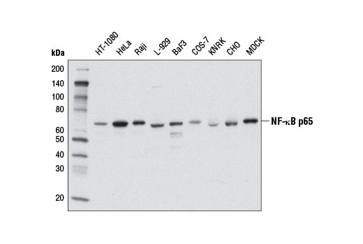
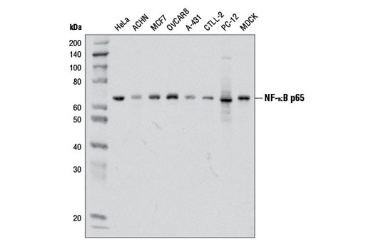
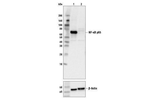
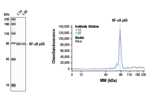
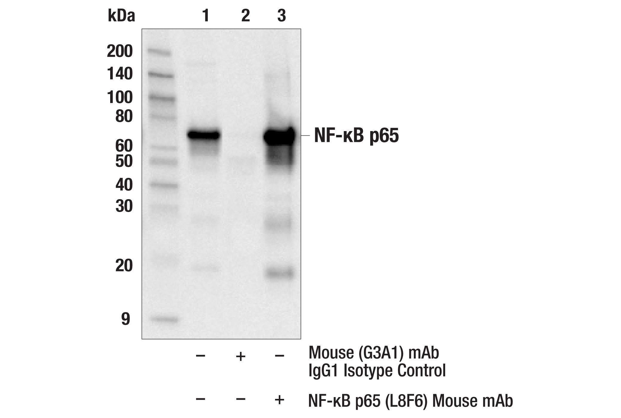
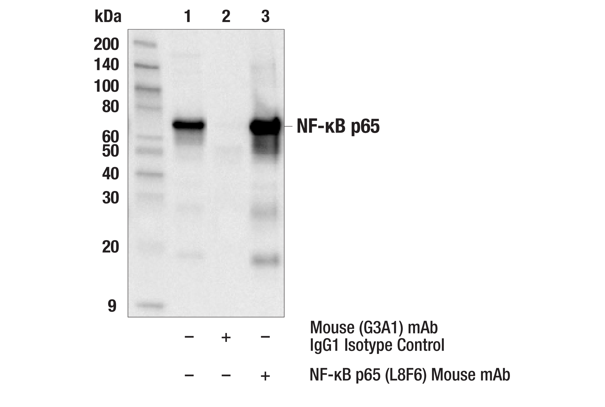
Western blot analysis of extracts from various cell lines using NF-κB p65 (L8F6) Mouse mAb.
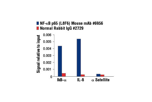
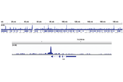
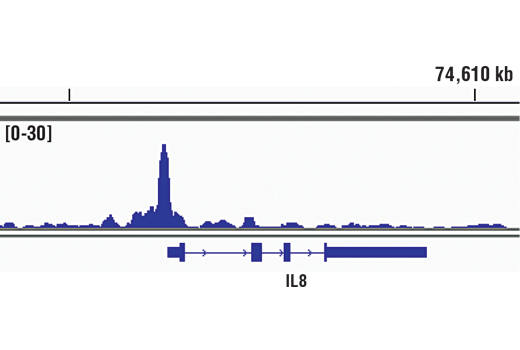
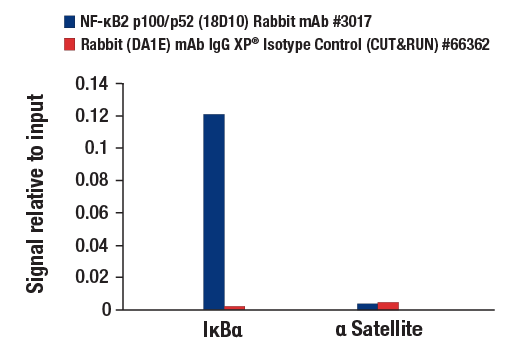
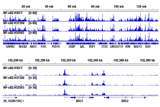
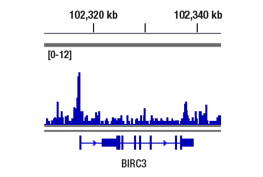
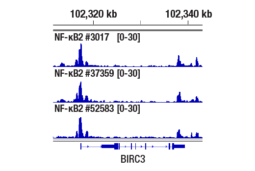
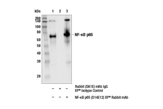
Chromatin immunoprecipitations were performed with cross-linked chromatin from 4 x 10⁶ HeLa cells treated with Human Tumor Necrosis Factor-α (hTNF-α) #8902 (30 ng/ml, 1 hr) and either 10 μl of NF-κB p65 (L8F6) Mouse mAb or 2 μl of Normal Rabbit IgG #2729 using SimpleChIP® Enzymatic Chromatin IP Kit (Magnetic Beads) #9003. The enriched DNA was quantified by real-time PCR using SimpleChIP® Human IκBα Promoter Primers #5552, human IL-8 promoter primers, and SimpleChIP® Human α Satellite Repeat Primers #4486. The amount of immunoprecipitated DNA in each sample is represented as signal relative to the total amount of input chromatin, which is equivalent to one.
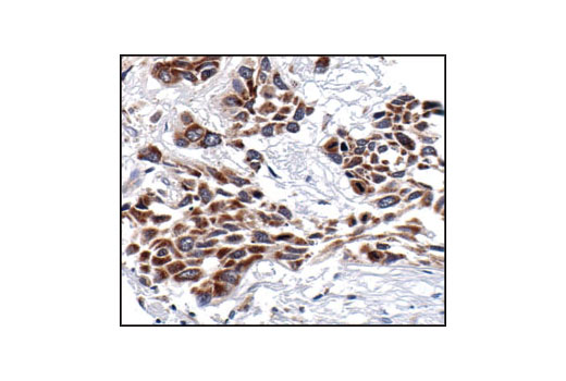
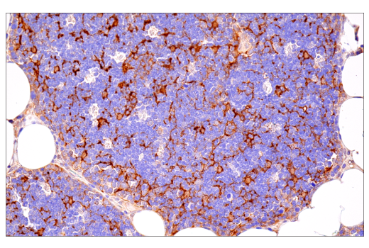
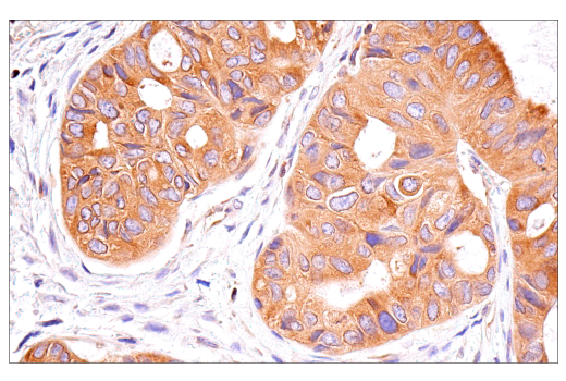
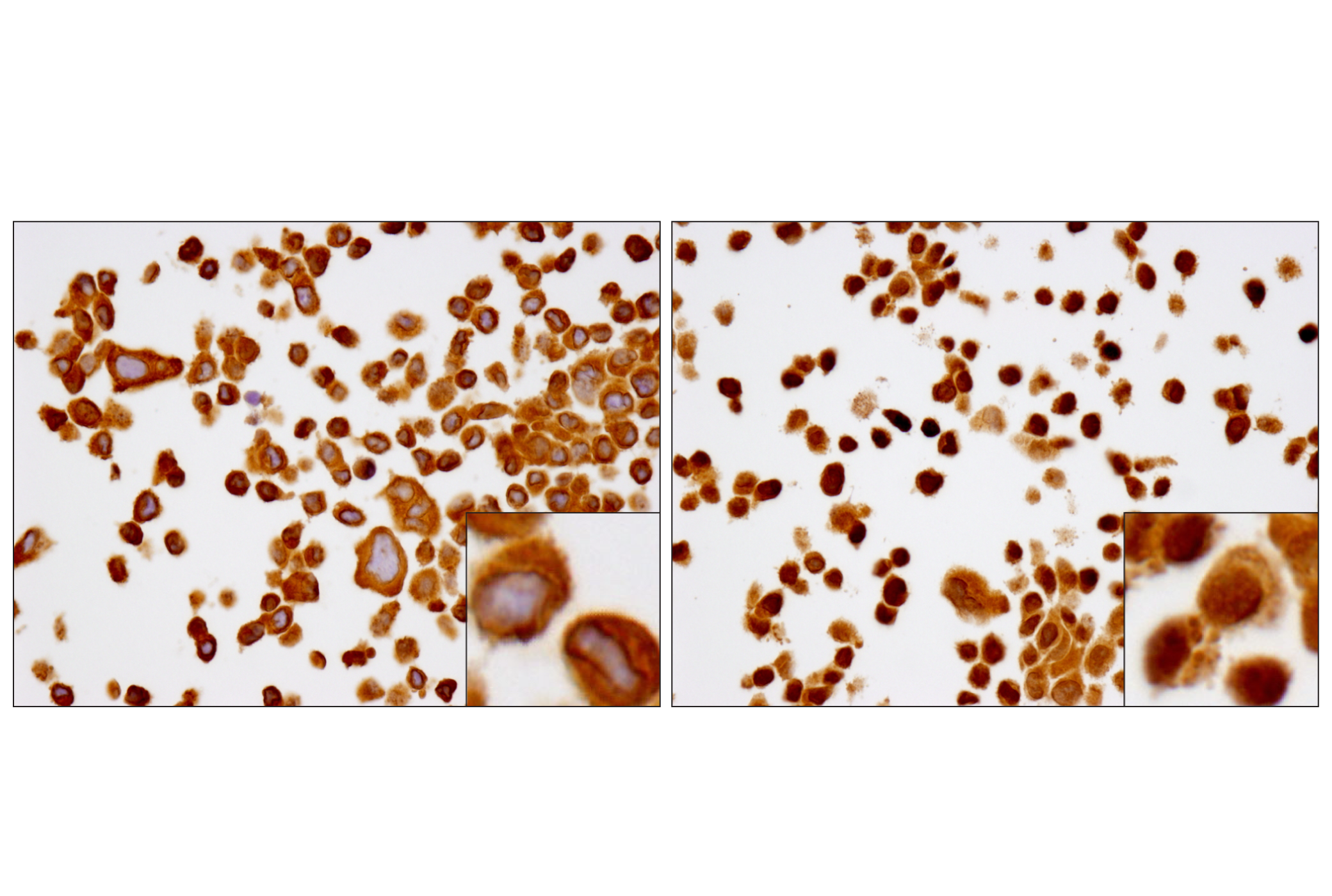
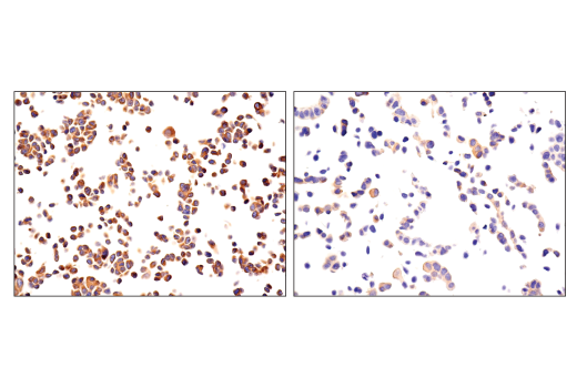
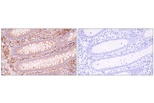
Immunohistochemical analysis of paraffin-embedded HeLa cell pellets, untreated (left) or treated with Human Tumor Necrosis Factor-α (hTNF-α) #8902 (right), using NF-κB p65 (L8F6) Mouse mAb.
Immunohistochemical analysis of paraffin-embedded OVCAR8 cell pellets treated with Human Tumor Necrosis Factor-α (hTNF-α) #8902 (left) or treated with SignalSilence® NF-κB p65 siRNA I #6261 (right), using NF-κB p65 (L8F6) Mouse mAb.
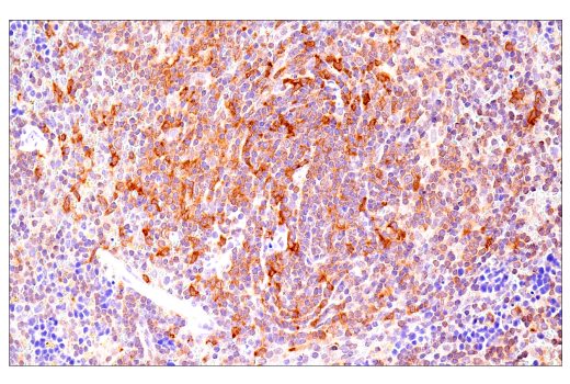
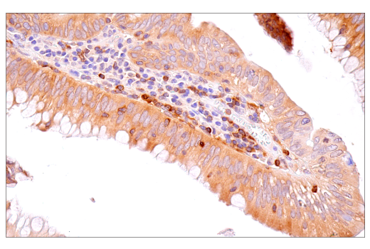
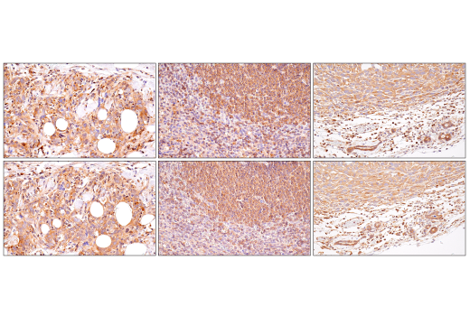
Immunohistochemical analysis of human chronic cholecystitis tissue using NF-κB p65 (L8F6) Mouse mAb.
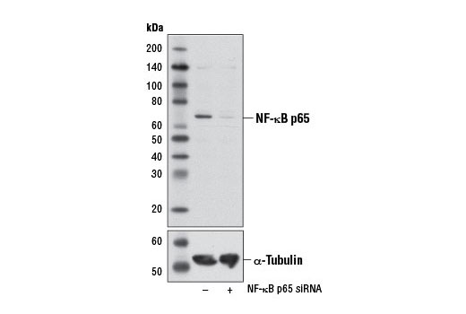
Western blot analysis of extracts from HeLa cells, transfected with 100 nM SignalSilence® Control siRNA (Unconjugated) #6568 (-) or SignalSilence® NF-κB p65 siRNA I #6261 (+), using NF-κB p65 (L8F6) Mouse mAb (upper) or α-Tubulin (11Η10) Rabbit mAb #2125 (lower). The NF-κB p65 (L8F6) Mouse mAb confirms silencing of NF-κB p65 expression, while the α-Tubulin (11Η10) Rabbit mAb is used as a loading control.
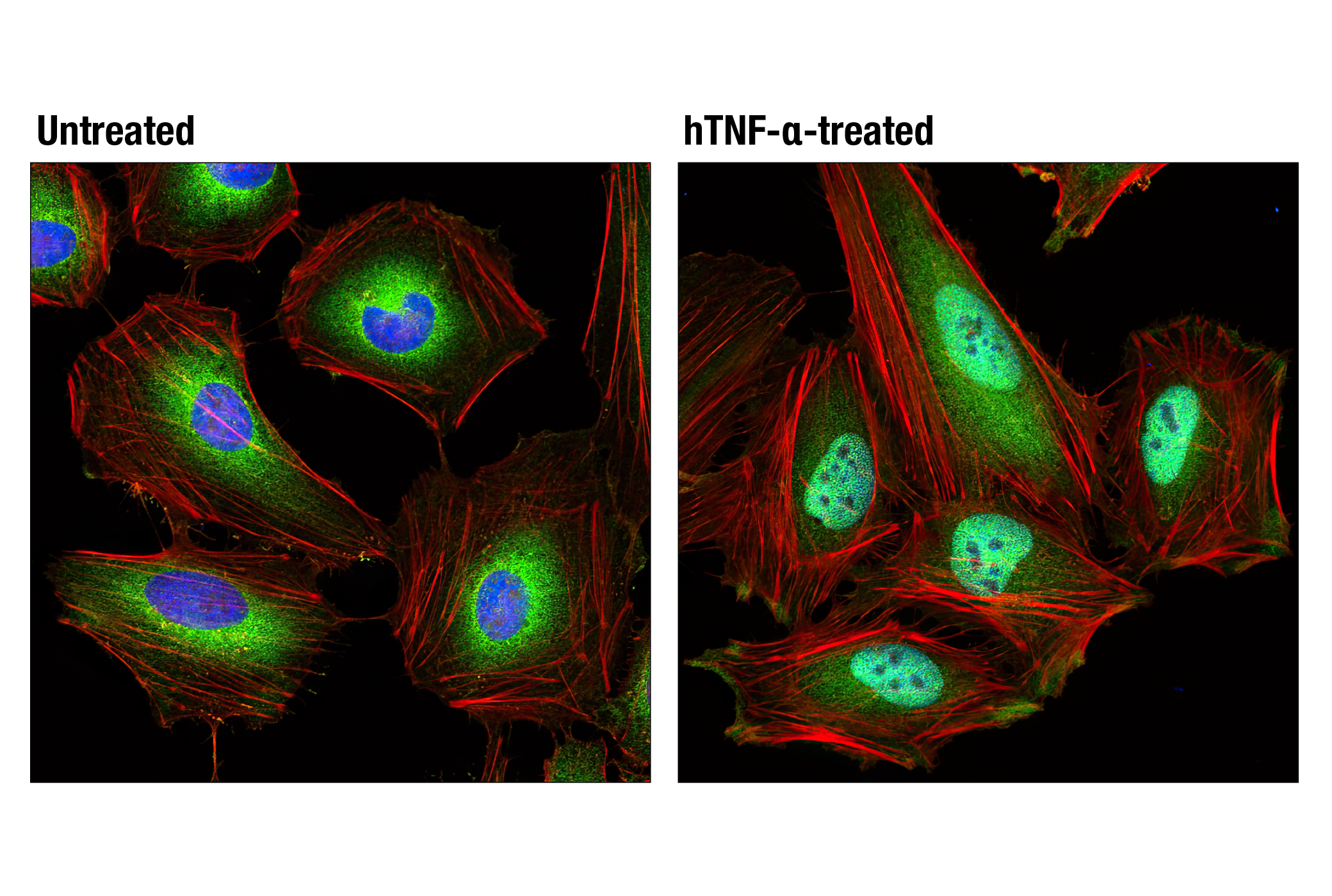
Confocal immunofluorescent analysis of HeLa cells, untreated (left) or treated with Human Tumor Necrosis Factor-α (hTNF-α) #8902 (20 ng/mL, 20 min; right), using NF-κB p65 (L8F6) Mouse mAb (green). Actin filaments were labeled with DY-554 phalloidin (red). Blue pseudocolor = DRAQ5® #4084 (fluorescent DNA dye).
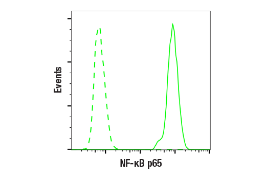
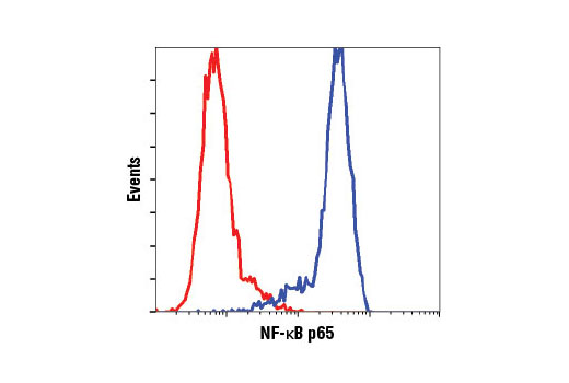
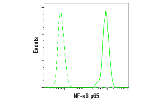
Flow cytometric analysis of HeLa cells using NF-κB p65 (L8F6) Mouse mAb (blue) compared to a nonspecific negative control antibody (red).
Confocal immunofluorescent analysis of HT-1080 cells, untreated (left) or treated with hTNF-α #8902 (20 ng/ml, 20 min) (right), using NF-κB p65 (D14E12) XP® Rabbit mAb (green). Actin filaments were labeled with DY-554 phalloidin (red). Blue pseudocolor = DRAQ5® #4084 (fluorescent DNA dye).
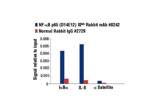
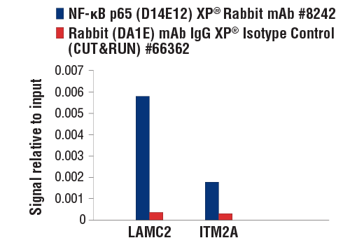
Chromatin immunoprecipitations were performed with cross-linked chromatin from 4 x 10⁶ HeLa cells treated with hTNF-α #8902 (30 ng/ml, 1 hr) and either 5 μl of NF-κB p65 (D14E12) XP® Rabbit mAb or 2 μl of Normal Rabbit IgG #2729 using SimpleChIP® Enzymatic Chromatin IP Kit (Magnetic Beads) #9003. The enriched DNA was quantified by Real-Time PCR using SimpleChIP® Human IκBα Promoter Primers #5552, human IL-8 promoter primers, and SimpleChIP® Human α Satellite Repeat Primers #4486. The amount of immunoprecipitated DNA in each sample is represented as signal relative to the total amount of input chromatin, which is equivalent to one.
Western blot analysis of extracts from various cell lines using NF-κB p65 (D14E12) XP® Rabbit mAb.
Western blot analysis of extracts from various cell lines using NF-κβ p65 (L8F6) Mouse mAb #6956.Western免疫印迹。用NF-κβ p65 (L8F6) Mouse mAb #6956研究各种细胞系的细胞提取液。
Western blot analysis of extracts from THP-1 cells, differentiated with TPA (#9905, 80 nM for 24 h) and treated with 1 μg/ml LPS for the indicated times, using NF-κβ1 p105 Antibody #4717.Western免疫印迹。用NF-κβ1 p105 Antibody #4717研究经TPA(#9905, 80 nM,24 h) 分化处理然后经过1 μg/ml LPS 处理一定时间的THP-1细胞的细胞提取液。
Flow cytometric analysis of HeLa cells using NF-κB p65 (D14E12) XP® Rabbit mAb (blue) compared to concentration matched Rabbit (DA1E) mAb IgG XP® Isotype Control #3900 (red).
Immunohistochemical analysis of paraffin-embedded human chronic cholecystitis using NF-κB p65 (D14E12) XP® Rabbit mAb.
Western blot analysis of extracts from various cell lines using NF-κB p65 (D14E12) XP® Rabbit mAb #8242.Western免疫印迹。用NF-κB p65 (D14E12) XP® Rabbit mAb #8242研究各种细胞系的细胞提取液。
Chromatin immunoprecipitations were performed with cross-linked chromatin from 4 x 10⁶ HeLa cells treated with Human Tumor Necrosis Factor-α (hTNF-α) #8902 (30 ng/ml) for 1 hour and either 10 μl of NF-κB1 p105/p50 (D7H5M) Rabbit mAb or 2 μl of Normal Rabbit IgG #2729 using SimpleChIP® Enzymatic Chromatin IP Kit (Magnetic Beads) #9003. The enriched DNA was quantified by Real-Time PCR using SimpleChIP® Human IκBα Promoter Primers #5552, human IAP2 promoter primers, and SimpleChIP® Human α Satellite Repeat Primers #4486. The amount of immunoprecipitated DNA in each sample is represented as signal relative to the total amount of input chromatin, which is equivalent to one.
Western blot analysis of extracts from various cell lines using NF-κB1 p105/p50 (D7H5M) Rabbit mAb.
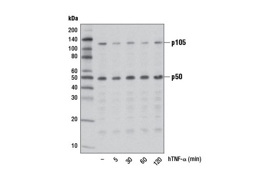
Western blot analysis of extracts from HeLa cells, untreated (-) or treated with Human Tumor Necrosis Factor-α (hTNF-α) #8902 (10 ng/ml) for the indicated times, using NF-κB1 p105/p50 (D7H5M) Rabbit mAb.

 危险品化学品经营许可证(不带存储) 许可证编号:沪(杨)应急管危经许[2022]202944(QY)
  营业执照(三证合一)